6EE0

Crystal Structure of SNX23 PX domain


Experimental Data Snapshot

  • Method: X-RAY DIFFRACTION
  • Resolution: 2.52 Å
  • R-Value Free: 0.247 
  • R-Value Work: 0.178 
  • R-Value Observed: 0.185 

wwPDB Validation   3D Report Full Report


This is version 1.3 of the entry. See complete history


Literature

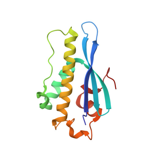
Classification of the human phox homology (PX) domains based on their phosphoinositide binding specificities.

Chandra, M.Chin, Y.K.Mas, C.Feathers, J.R.Paul, B.Datta, S.Chen, K.E.Jia, X.Yang, Z.Norwood, S.J.Mohanty, B.Bugarcic, A.Teasdale, R.D.Henne, W.M.Mobli, M.Collins, B.M.

(2019) Nat Commun 10: 1528-1528

  • DOI: https://doi.org/10.1038/s41467-019-09355-y
  • Primary Citation of Related Structures:  
    5WOE, 6ECM, 6EDX, 6EE0, 6MBI

  • PubMed Abstract: 

    Phox homology (PX) domains are membrane interacting domains that bind to phosphatidylinositol phospholipids or phosphoinositides, markers of organelle identity in the endocytic system. Although many PX domains bind the canonical endosome-enriched lipid PtdIns3P, others interact with alternative phosphoinositides, and a precise understanding of how these specificities arise has remained elusive. Here we systematically screen all human PX domains for their phospholipid preferences using liposome binding assays, biolayer interferometry and isothermal titration calorimetry. These analyses define four distinct classes of human PX domains that either bind specifically to PtdIns3P, non-specifically to various di- and tri-phosphorylated phosphoinositides, bind both PtdIns3P and other phosphoinositides, or associate with none of the lipids tested. A comprehensive evaluation of PX domain structures reveals two distinct binding sites that explain these specificities, providing a basis for defining and predicting the functional membrane interactions of the entire PX domain protein family.


  • Organizational Affiliation

    Institute for Molecular Bioscience, The University of Queensland, St. Lucia, QLD, 4072, Australia.


Macromolecules
Find similar proteins by:  (by identity cutoff)  |  3D Structure
Entity ID: 1
MoleculeChains Sequence LengthOrganismDetailsImage
Kinesin-like protein KIF16B
A, B, C
133Homo sapiensMutation(s): 0 
Gene Names: KIF16BC20orf23KIAA1590SNX23
Membrane Entity: Yes 
UniProt & NIH Common Fund Data Resources
Find proteins for Q96L93 (Homo sapiens)
Explore Q96L93 
Go to UniProtKB:  Q96L93
PHAROS:  Q96L93
GTEx:  ENSG00000089177 
Entity Groups  
Sequence Clusters30% Identity50% Identity70% Identity90% Identity95% Identity100% Identity
UniProt GroupQ96L93
Sequence Annotations
Expand
  • Reference Sequence
Experimental Data & Validation

Experimental Data

  • Method: X-RAY DIFFRACTION
  • Resolution: 2.52 Å
  • R-Value Free: 0.247 
  • R-Value Work: 0.178 
  • R-Value Observed: 0.185 
  • Space Group: P 1 21 1
Unit Cell:
Length ( Å )Angle ( ˚ )
a = 46.783α = 90
b = 92.836β = 91.29
c = 47.039γ = 90
Software Package:
Software NamePurpose
PHENIXrefinement
XDSdata reduction
SCALAdata scaling
PHASERphasing

Structure Validation

View Full Validation Report


Entry History & Funding Information

Deposition Data


Funding OrganizationLocationGrant Number
Australian Research Council (ARC)AustraliaDP160101743
National Health and Medical Research Council (NHMRC, Australia)AustraliaAPP1099114
National Health and Medical Research Council (NHMRC, Australia)AustraliaAPP1136021

Revision History  (Full details and data files)

  • Version 1.0: 2018-08-22
    Type: Initial release
  • Version 1.1: 2019-04-17
    Changes: Data collection, Database references
  • Version 1.2: 2020-01-01
    Changes: Author supporting evidence
  • Version 1.3: 2023-10-11
    Changes: Data collection, Database references, Refinement description