6DSV

Bst DNA polymerase I post-chemistry (n+2) structure


Experimental Data Snapshot

  • Method: X-RAY DIFFRACTION
  • Resolution: 1.99 Å
  • R-Value Free: 0.228 
  • R-Value Work: 0.193 
  • R-Value Observed: 0.194 

wwPDB Validation   3D Report Full Report


This is version 1.2 of the entry. See complete history


Literature

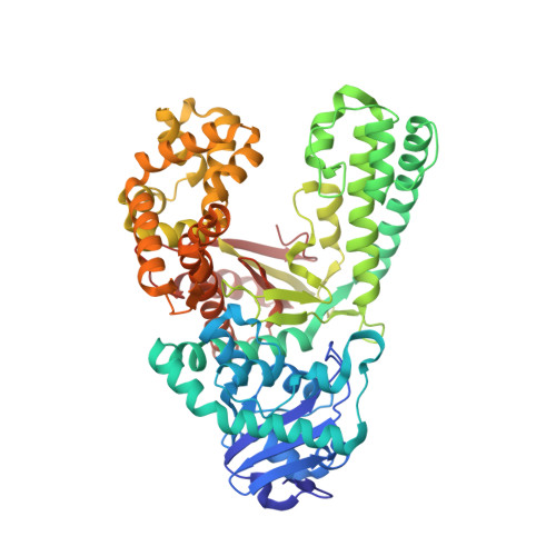
Crystal structures of DNA polymerase I capture novel intermediates in the DNA synthesis pathway.

Chim, N.Jackson, L.N.Trinh, A.M.Chaput, J.C.

(2018) Elife 7

  • DOI: https://doi.org/10.7554/eLife.40444
  • Primary Citation of Related Structures:  
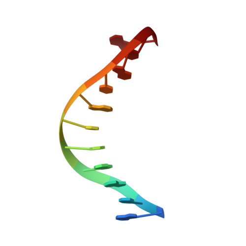
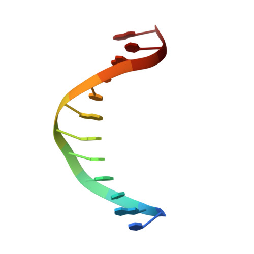
    6DSU, 6DSV, 6DSW, 6DSX, 6DSY

  • PubMed Abstract: 

    High resolution crystal structures of DNA polymerase intermediates are needed to study the mechanism of DNA synthesis in cells. Here we report five crystal structures of DNA polymerase I that capture new conformations for the polymerase translocation and nucleotide pre-insertion steps in the DNA synthesis pathway. We suggest that these new structures, along with previously solved structures, highlight the dynamic nature of the finger subdomain in the enzyme active site.


  • Organizational Affiliation

    Departments of Pharmaceutical Sciences, University of California, Irvine, California.


Macromolecules

Find similar proteins by:  (by identity cutoff)  |  3D Structure
Entity ID: 2
MoleculeChains Sequence LengthOrganismDetailsImage
DNA polymerase IB [auth A]580Geobacillus stearothermophilusMutation(s): 0 
Gene Names: polA
EC: 2.7.7.7
UniProt
Find proteins for E1C9K5 (Geobacillus stearothermophilus)
Explore E1C9K5 
Go to UniProtKB:  E1C9K5
Entity Groups  
Sequence Clusters30% Identity50% Identity70% Identity90% Identity95% Identity100% Identity
UniProt GroupE1C9K5
Sequence Annotations
Expand
  • Reference Sequence

Find similar nucleic acids by:  Sequence   |   3D Structure  

Entity ID: 1
MoleculeChains LengthOrganismImage
DNA (5'-D(*GP*CP*GP*AP*TP*CP*AP*CP*GP*TP*A)-3')A [auth P]11synthetic construct
Sequence Annotations
Expand
  • Reference Sequence

Find similar nucleic acids by:  Sequence   |   3D Structure  

Entity ID: 3
MoleculeChains LengthOrganismImage
DNA (5'-D(P*TP*AP*CP*GP*TP*GP*AP*TP*CP*GP*CP*A)-3')C [auth T]12synthetic construct
Sequence Annotations
Expand
  • Reference Sequence
Experimental Data & Validation

Experimental Data

  • Method: X-RAY DIFFRACTION
  • Resolution: 1.99 Å
  • R-Value Free: 0.228 
  • R-Value Work: 0.193 
  • R-Value Observed: 0.194 
  • Space Group: P 21 21 21
Unit Cell:
Length ( Å )Angle ( ˚ )
a = 87.082α = 90
b = 93.069β = 90
c = 104.736γ = 90
Software Package:
Software NamePurpose
PHENIXrefinement
autoXDSdata reduction
autoXDSdata scaling
PHASERphasing

Structure Validation

View Full Validation Report


Entry History & Funding Information

Deposition Data


Funding OrganizationLocationGrant Number
National Science Foundation (NSF, United States)United StatesMCB: 1607111

Revision History  (Full details and data files)

  • Version 1.0: 2018-10-31
    Type: Initial release
  • Version 1.1: 2019-11-27
    Changes: Author supporting evidence
  • Version 1.2: 2024-03-13
    Changes: Data collection, Database references