6DG2

Antigen Binding Fragment of the Pan-ebolavirus Monoclonal Antibody 6D6


Experimental Data Snapshot

  • Method: X-RAY DIFFRACTION
  • Resolution: 1.96 Å
  • R-Value Free: 0.235 
  • R-Value Work: 0.191 
  • R-Value Observed: 0.192 

wwPDB Validation   3D Report Full Report


This is version 1.3 of the entry. See complete history


Literature

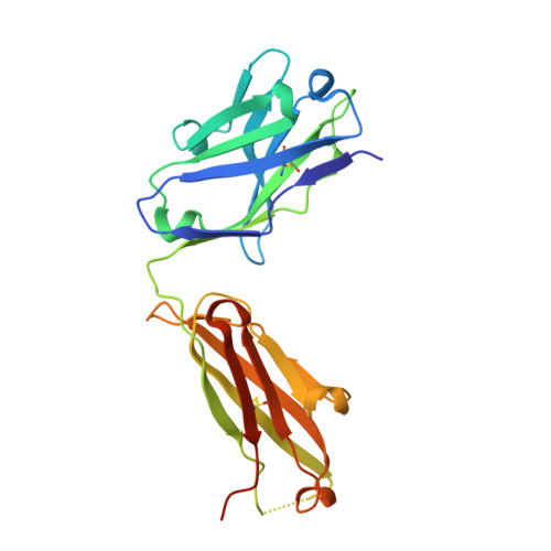
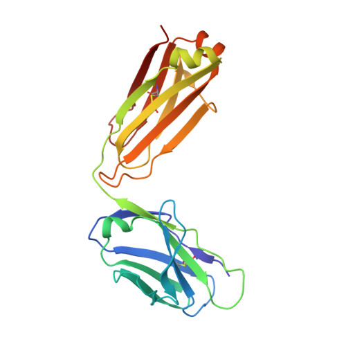
Structural Characterization of Pan-Ebolavirus Antibody 6D6 Targeting the Fusion Peptide of the Surface Glycoprotein.

Milligan, J.C.Parekh, D.V.Fuller, K.M.Igarashi, M.Takada, A.Saphire, E.O.

(2019) J Infect Dis 219: 415-419

  • DOI: https://doi.org/10.1093/infdis/jiy532
  • Primary Citation of Related Structures:  
    6DG2

  • PubMed Abstract: 

    Ebola virus infection causes severe disease in humans and represents a global health threat. Candidates for immunotherapeutics and vaccines have shown promise in clinical trials, although they are ineffective against other members of the Ebolavirus genus that also cause periodic, lethal outbreaks. In this study, we present a crystal structure of a pan-ebolavirus antibody, 6D6, as well as single-particle electron microscopy reconstructions of 6D6 in complex with Ebola and Bundibugyo virus glycoproteins. 6D6 binds to the conserved glycoprotein fusion peptide, implicating it as a site of immune vulnerability that could be exploited to reliably elicit a pan-ebolavirus neutralizing antibody response.


  • Organizational Affiliation

    Department of Immunology and Microbiology, La Jolla, California.


Macromolecules
Find similar proteins by:  (by identity cutoff)  |  3D Structure
Entity ID: 1
MoleculeChains Sequence LengthOrganismDetailsImage
6D6 Fab heavy chain
A, C
230Mus musculusMutation(s): 0 
Entity Groups  
Sequence Clusters30% Identity50% Identity70% Identity90% Identity95% Identity100% Identity
Sequence Annotations
Expand
  • Reference Sequence
Find similar proteins by:  (by identity cutoff)  |  3D Structure
Entity ID: 2
MoleculeChains Sequence LengthOrganismDetailsImage
6D6 Fab light chain
B, D
215Mus musculusMutation(s): 0 
Entity Groups  
Sequence Clusters30% Identity50% Identity70% Identity90% Identity95% Identity100% Identity
Sequence Annotations
Expand
  • Reference Sequence
Experimental Data & Validation

Experimental Data

  • Method: X-RAY DIFFRACTION
  • Resolution: 1.96 Å
  • R-Value Free: 0.235 
  • R-Value Work: 0.191 
  • R-Value Observed: 0.192 
  • Space Group: P 1 21 1
Unit Cell:
Length ( Å )Angle ( ˚ )
a = 70.226α = 90
b = 72.038β = 104.2
c = 107.149γ = 90
Software Package:
Software NamePurpose
PHENIXrefinement
MOSFLMdata reduction
Aimlessdata scaling
PHENIXphasing

Structure Validation

View Full Validation Report


Entry History & Funding Information

Deposition Data


Funding OrganizationLocationGrant Number
National Institutes of Health/National Institute Of Allergy and Infectious Diseases (NIH/NIAID)United States5U19AI109762-05

Revision History  (Full details and data files)

  • Version 1.0: 2018-09-26
    Type: Initial release
  • Version 1.1: 2019-01-23
    Changes: Data collection, Database references
  • Version 1.2: 2019-12-18
    Changes: Author supporting evidence
  • Version 1.3: 2023-10-11
    Changes: Data collection, Database references, Refinement description