6DA8

Human CYP3A4 bound to an inhibitor


Experimental Data Snapshot

  • Method: X-RAY DIFFRACTION
  • Resolution: 2.80 Å
  • R-Value Free: 0.291 
  • R-Value Work: 0.216 
  • R-Value Observed: 0.220 

wwPDB Validation   3D Report Full Report


Ligand Structure Quality Assessment 


This is version 1.4 of the entry. See complete history


Literature

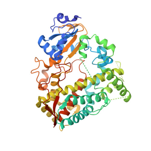
Structure-Activity Relationships of Rationally Designed Ritonavir Analogues: Impact of Side-Group Stereochemistry, Headgroup Spacing, and Backbone Composition on the Interaction with CYP3A4.

Samuels, E.R.Sevrioukova, I.

(2019) Biochemistry 58: 2077-2087

  • DOI: https://doi.org/10.1021/acs.biochem.9b00156
  • Primary Citation of Related Structures:  
    6DA2, 6DA3, 6DA5, 6DA8, 6DAA, 6DAB, 6DAC, 6DAG, 6DAJ, 6DAL

  • PubMed Abstract: 

    In a continuing effort to identify structural attributes required for strong binding and potent inhibition of human drug-metabolizing CYP3A4, we designed ten ritonavir-like analogues differing in the side-group stereochemistry, backbone atomic composition, and headgroup spacing. All analogues had pyridine and tert-butyloxycarbonyl (Boc) as the heme-ligating head and tail groups, respectively, phenyl side groups, and either a methyl- or ethyl-pyridyl linker. Each linker subseries had S/ R, R/ S, R/ R, and S/S side-group conformers (4a-d and 4e-h, respectively), and one S/S stereoisomer with the backbone S-to-N-heteroatom substitution (6a and 6b). To elucidate structure-activity relationships, ligand-dependent changes in optical spectra, dissociation constant ( K s ), inhibitory potency (IC 50 ), thermostability, and heme ligation and reduction kinetics were analyzed. Comparison of the subseries and individual compounds showed that CYP3A4 only weakly discriminates between side-group configurations, associates more tightly with the pyridyl-ethyl-linker analogues, and strongly disfavors the N-containing backbone. K s and IC 50 for the pyridyl-ethyl R/ R conformer, 4g, were the lowest and close to those for ritonavir: 0.04 and 0.31 μM versus 0.02 and 0.13 μM, respectively. Determination of the X-ray structures of the inhibitory complexes was critical for experimental data interpretation, especially for the uniquely oriented 4a and 4e. Based on structural analysis, we conclude that, for this series of analogues, the ligand-mediated interactions near the heme are dominant and define the binding mode and that fine-tuning of these interactions as well as the backbone spacing could further improve the affinity and inhibitory strength.


Macromolecules
Find similar proteins by:  (by identity cutoff)  |  3D Structure
Entity ID: 1
MoleculeChains Sequence LengthOrganismDetailsImage
Cytochrome P450 3A4487Homo sapiensMutation(s): 0 
Gene Names: CYP3A4CYP3A3
EC: 1.14.14 (PDB Primary Data), 1.14.14.56 (PDB Primary Data), 1.14.14.55 (PDB Primary Data)
UniProt & NIH Common Fund Data Resources
Find proteins for P08684 (Homo sapiens)
Explore P08684 
Go to UniProtKB:  P08684
PHAROS:  P08684
GTEx:  ENSG00000160868 
Entity Groups  
Sequence Clusters30% Identity50% Identity70% Identity90% Identity95% Identity100% Identity
UniProt GroupP08684
Sequence Annotations
Expand
  • Reference Sequence
Small Molecules
Ligands 2 Unique
IDChains Name / Formula / InChI Key2D Diagram3D Interactions
HEM
Query on HEM

Download Ideal Coordinates CCD File 
B [auth A]PROTOPORPHYRIN IX CONTAINING FE
C34 H32 Fe N4 O4
KABFMIBPWCXCRK-RGGAHWMASA-L
G0J
Query on G0J

Download Ideal Coordinates CCD File 
C [auth A]Nalpha-{(2S)-2-[(tert-butoxycarbonyl)amino]-3-phenylpropyl}-N-[(pyridin-3-yl)methyl]-D-phenylalaninamide
C29 H36 N4 O3
RLXQYSRXFSZPNG-IZZNHLLZSA-N
Experimental Data & Validation

Experimental Data

  • Method: X-RAY DIFFRACTION
  • Resolution: 2.80 Å
  • R-Value Free: 0.291 
  • R-Value Work: 0.216 
  • R-Value Observed: 0.220 
  • Space Group: I 2 2 2
Unit Cell:
Length ( Å )Angle ( ˚ )
a = 76.25α = 90
b = 101.68β = 90
c = 126.24γ = 90
Software Package:
Software NamePurpose
PHENIXrefinement
MOSFLMdata reduction
SCALAdata scaling
PHASERphasing

Structure Validation

View Full Validation Report


Ligand Structure Quality Assessment 


Entry History & Funding Information

Deposition Data


Funding OrganizationLocationGrant Number
National Institutes of Health/National Institute of Environmental Health Sciences (NIH/NIEHS)United StatesES025767

Revision History  (Full details and data files)

  • Version 1.0: 2019-04-03
    Type: Initial release
  • Version 1.1: 2019-05-01
    Changes: Data collection, Database references
  • Version 1.2: 2019-06-12
    Changes: Data collection, Database references
  • Version 1.3: 2019-12-18
    Changes: Author supporting evidence
  • Version 1.4: 2023-10-04
    Changes: Data collection, Database references, Refinement description