6D1U

Crystal structure of the human CLR:RAMP1 extracellular domain heterodimer in complex with adrenomedullin 2/intermedin


Experimental Data Snapshot

  • Method: X-RAY DIFFRACTION
  • Resolution: 2.05 Å
  • R-Value Free: 0.223 
  • R-Value Work: 0.194 
  • R-Value Observed: 0.195 

wwPDB Validation   3D Report Full Report


This is version 2.1 of the entry. See complete history


Literature

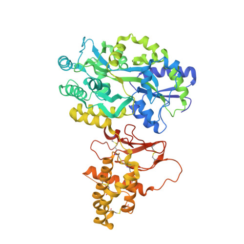
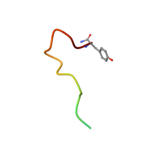
Structure-function analyses reveal a triple beta-turn receptor-bound conformation of adrenomedullin 2/intermedin and enable peptide antagonist design.

Roehrkasse, A.M.Booe, J.M.Lee, S.M.Warner, M.L.Pioszak, A.A.

(2018) J Biol Chem 293: 15840-15854

  • DOI: https://doi.org/10.1074/jbc.RA118.005062
  • Primary Citation of Related Structures:  
    6D1U

  • PubMed Abstract: 

    The cardioprotective vasodilator peptide adrenomedullin 2/intermedin (AM2/IMD) and the related adrenomedullin (AM) and calcitonin gene-related peptide (CGRP) signal through three heterodimeric receptors comprising the calcitonin receptor-like class B G protein-coupled receptor (CLR) and a variable receptor activity-modifying protein (RAMP1, -2, or -3) that determines ligand selectivity. The CGRP receptor (RAMP1:CLR) favors CGRP binding, whereas the AM 1 (RAMP2:CLR) and AM 2 (RAMP3:CLR) receptors favor AM binding. How AM2/IMD binds the receptors and how RAMPs modulate its binding is unknown. Here, we show that AM2/IMD binds the three purified RAMP-CLR extracellular domain (ECD) complexes with a selectivity profile that is distinct from those of CGRP and AM. AM2/IMD bound all three ECD complexes but preferred the CGRP and AM 2 receptor complexes. A 2.05 Å resolution crystal structure of an AM2/IMD antagonist fragment-bound RAMP1-CLR ECD complex revealed that AM2/IMD binds the complex through a unique triple β-turn conformation that was confirmed by peptide and receptor mutagenesis. Comparisons of the receptor-bound conformations of AM2/IMD, AM, and a high-affinity CGRP analog revealed differences that may have implications for biased signaling. Guided by the structure, enhanced-affinity AM2/IMD antagonist variants were developed, including one that discriminates the AM 1 and AM 2 receptors with ∼40-fold difference in affinities and one stabilized by an intramolecular disulfide bond. These results reveal differences in how the three peptides engage the receptors, inform development of AM2/IMD-based pharmacological tools and therapeutics, and provide insights into RAMP modulation of receptor pharmacology.


  • Organizational Affiliation

    From the Department of Biochemistry and Molecular Biology, University of Oklahoma Health Sciences Center, Oklahoma City, Oklahoma 73104.


Macromolecules
Find similar proteins by:  (by identity cutoff)  |  3D Structure
Entity ID: 1
MoleculeChains Sequence LengthOrganismDetailsImage
Maltose-binding periplasmic protein,Receptor activity-modifying protein 1,Calcitonin gene-related peptide type 1 receptor
A, B, C
593Escherichia coli O157:H7Homo sapiensMutation(s): 0 
Gene Names: malEZ5632ECs5017RAMP1CALCRLCGRPR
UniProt & NIH Common Fund Data Resources
Find proteins for P0AEX9 (Escherichia coli (strain K12))
Explore P0AEX9 
Go to UniProtKB:  P0AEX9
Find proteins for O60894 (Homo sapiens)
Explore O60894 
Go to UniProtKB:  O60894
PHAROS:  O60894
GTEx:  ENSG00000132329 
Find proteins for Q16602 (Homo sapiens)
Explore Q16602 
Go to UniProtKB:  Q16602
PHAROS:  Q16602
GTEx:  ENSG00000064989 
Entity Groups  
Sequence Clusters30% Identity50% Identity70% Identity90% Identity95% Identity100% Identity
UniProt GroupsP0AEX9Q16602O60894
Sequence Annotations
Expand
  • Reference Sequence

Find similar proteins by:  Sequence   |   3D Structure  

Entity ID: 2
MoleculeChains Sequence LengthOrganismDetailsImage
ADM2
D, E, F
20Homo sapiensMutation(s): 0 
UniProt & NIH Common Fund Data Resources
Find proteins for Q7Z4H4 (Homo sapiens)
Explore Q7Z4H4 
Go to UniProtKB:  Q7Z4H4
PHAROS:  Q7Z4H4
GTEx:  ENSG00000128165 
Entity Groups  
Sequence Clusters30% Identity50% Identity70% Identity90% Identity95% Identity100% Identity
UniProt GroupQ7Z4H4
Sequence Annotations
Expand
  • Reference Sequence
Oligosaccharides

Help

Entity ID: 3
MoleculeChains Length2D Diagram Glycosylation3D Interactions
alpha-D-glucopyranose-(1-4)-alpha-D-glucopyranose
G, H, I
2N/A
Glycosylation Resources
GlyTouCan:  G07411ON
GlyCosmos:  G07411ON
Biologically Interesting Molecules (External Reference) 1 Unique
Experimental Data & Validation

Experimental Data

  • Method: X-RAY DIFFRACTION
  • Resolution: 2.05 Å
  • R-Value Free: 0.223 
  • R-Value Work: 0.194 
  • R-Value Observed: 0.195 
  • Space Group: C 1 2 1
Unit Cell:
Length ( Å )Angle ( ˚ )
a = 172.295α = 90
b = 106.041β = 122.53
c = 136.911γ = 90
Software Package:
Software NamePurpose
HKL-2000data scaling
REFMACrefinement
PDB_EXTRACTdata extraction
HKL-2000data reduction
PHASERphasing

Structure Validation

View Full Validation Report


Entry History & Funding Information

Deposition Data


Funding OrganizationLocationGrant Number
National Institutes of Health/National Institute of General Medical Sciences (NIH/NIGMS)United StatesR01GM104251

Revision History  (Full details and data files)

  • Version 1.0: 2018-09-05
    Type: Initial release
  • Version 1.1: 2018-10-24
    Changes: Data collection, Database references
  • Version 1.2: 2020-01-01
    Changes: Author supporting evidence
  • Version 2.0: 2020-07-29
    Type: Remediation
    Reason: Carbohydrate remediation
    Changes: Atomic model, Data collection, Derived calculations, Non-polymer description, Structure summary
  • Version 2.1: 2023-10-04
    Changes: Data collection, Database references, Refinement description, Structure summary