6BX8

Human Mesotrypsin (PRSS3) Complexed with Tissue Factor Pathway Inhibitor Variant (TFPI1-KD1-K15R-I17C-I34C)


Experimental Data Snapshot

  • Method: X-RAY DIFFRACTION
  • Resolution: 1.98 Å
  • R-Value Free: 0.221 
  • R-Value Work: 0.192 
  • R-Value Observed: 0.194 

wwPDB Validation   3D Report Full Report


This is version 1.4 of the entry. See complete history


Literature

Disulfide engineering of human Kunitz-type serine protease inhibitors enhances proteolytic stability and target affinity toward mesotrypsin.

Cohen, I.Coban, M.Shahar, A.Sankaran, B.Hockla, A.Lacham, S.Caulfield, T.R.Radisky, E.S.Papo, N.

(2019) J Biol Chem 294: 5105-5120

  • DOI: https://doi.org/10.1074/jbc.RA118.007292
  • Primary Citation of Related Structures:  
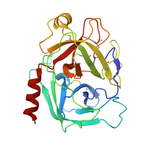
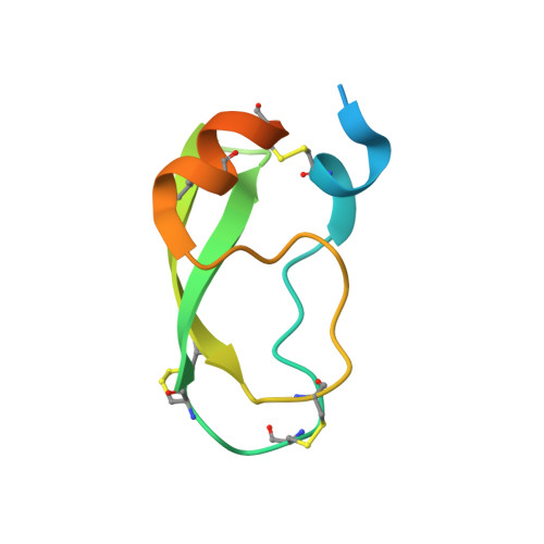
    6BX8, 6HAR

  • PubMed Abstract: 

    Serine protease inhibitors of the Kunitz-bovine pancreatic trypsin inhibitor (BPTI) family are ubiquitous biological regulators of proteolysis. These small proteins are resistant to proteolysis, but can be slowly cleaved within the protease-binding loop by target proteases, thereby compromising their activity. For the human protease mesotrypsin, this cleavage is especially rapid. Here, we aimed to stabilize the Kunitz domain structure against proteolysis through disulfide engineering. Substitution within the Kunitz inhibitor domain of the amyloid precursor protein (APPI) that incorporated a new disulfide bond between residues 17 and 34 reduced proteolysis by mesotrypsin 74-fold. Similar disulfide engineering of tissue factor pathway inhibitor-1 Kunitz domain 1 ( KD1 TFPI1) and bikunin Kunitz domain 2 ( KD2 bikunin) likewise stabilized these inhibitors against mesotrypsin proteolysis 17- and 6.6-fold, respectively. Crystal structures of disulfide-engineered APPI and KD1 TFPI1 variants in a complex with mesotrypsin at 1.5 and 2.0 Å resolution, respectively, confirmed the formation of well-ordered disulfide bonds positioned to stabilize the binding loop. Long all-atom molecular dynamics simulations of disulfide-engineered Kunitz domains and their complexes with mesotrypsin revealed conformational stabilization of the primed side of the inhibitor-binding loop by the engineered disulfide, along with global suppression of conformational dynamics in the Kunitz domain. Our findings suggest that the Cys-17-Cys-34 disulfide slows proteolysis by dampening conformational fluctuations in the binding loop and minimizing motion at the enzyme-inhibitor interface. The generalizable approach developed here for the stabilization against proteolysis of Kunitz domains, which can serve as important scaffolds for therapeutics, may thus find applications in drug development.


  • Organizational Affiliation

    From the Avram and Stella Goldstein-Goren Department of Biotechnology Engineering and the National Institute of Biotechnology in the Negev, Ben-Gurion University of the Negev, Beer-Sheva 84105, Israel.


Macromolecules
Find similar proteins by:  (by identity cutoff)  |  3D Structure
Entity ID: 1
MoleculeChains Sequence LengthOrganismDetailsImage
Trypsin-3
A, C, E, G
224Homo sapiensMutation(s): 1 
Gene Names: PRSS3PRSS4TRY3TRY4
EC: 3.4.21.4
UniProt & NIH Common Fund Data Resources
Find proteins for P35030 (Homo sapiens)
Explore P35030 
Go to UniProtKB:  P35030
PHAROS:  P35030
GTEx:  ENSG00000010438 
Entity Groups  
Sequence Clusters30% Identity50% Identity70% Identity90% Identity95% Identity100% Identity
UniProt GroupP35030
Sequence Annotations
Expand
  • Reference Sequence
Find similar proteins by:  (by identity cutoff)  |  3D Structure
Entity ID: 2
MoleculeChains Sequence LengthOrganismDetailsImage
Tissue factor pathway inhibitor
B, D, F, H
80Homo sapiensMutation(s): 3 
Gene Names: TFPILACITFPI1
UniProt & NIH Common Fund Data Resources
Find proteins for P10646 (Homo sapiens)
Explore P10646 
Go to UniProtKB:  P10646
PHAROS:  P10646
GTEx:  ENSG00000003436 
Entity Groups  
Sequence Clusters30% Identity50% Identity70% Identity90% Identity95% Identity100% Identity
UniProt GroupP10646
Sequence Annotations
Expand
  • Reference Sequence
Experimental Data & Validation

Experimental Data

  • Method: X-RAY DIFFRACTION
  • Resolution: 1.98 Å
  • R-Value Free: 0.221 
  • R-Value Work: 0.192 
  • R-Value Observed: 0.194 
  • Space Group: P 1
Unit Cell:
Length ( Å )Angle ( ˚ )
a = 35.137α = 94.81
b = 87.199β = 93.03
c = 90.84γ = 92.46
Software Package:
Software NamePurpose
PHENIXrefinement
Aimlessdata scaling
MOLREPphasing
PDB_EXTRACTdata extraction
DIALSdata reduction

Structure Validation

View Full Validation Report


Entry History & Funding Information

Deposition Data


Funding OrganizationLocationGrant Number
National Institutes of Health/National Library of Medicine (NIH/NLM)United StatesR01CA154387
United States - Israel Binational Science Foundation (BSF)IsraelBSF2015134

Revision History  (Full details and data files)

  • Version 1.0: 2019-02-06
    Type: Initial release
  • Version 1.1: 2019-02-13
    Changes: Data collection, Database references
  • Version 1.2: 2019-04-10
    Changes: Data collection, Database references
  • Version 1.3: 2019-12-18
    Changes: Author supporting evidence
  • Version 1.4: 2023-10-04
    Changes: Data collection, Database references, Refinement description