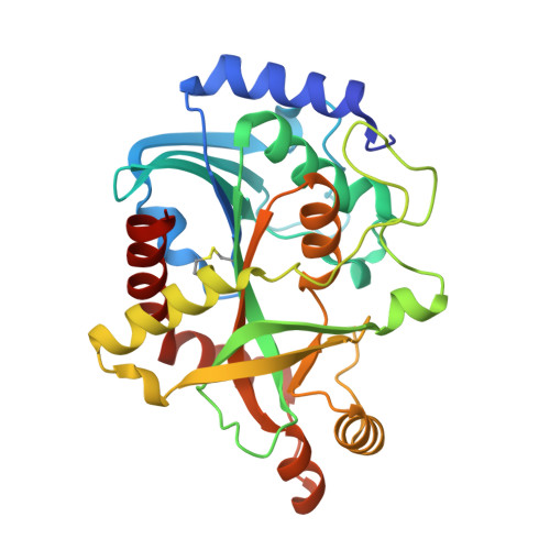
Crystal Structure of Purine Nucleoside Phosphorylase Isoform 2 from Schistosoma mansoni in complex with 1,2,5-trimethyl-1H-pyrrole-3-carboxylic acid
Faheem, M., Neto, J.B., Collins, P., Pearce, N.M., Valadares, N.F., Bird, L., Pereira, H.M., Delft, F.V., Barbosa, J.A.R.G.To be published.