6B2D

Crystal structure of fluoride channel Fluc Ec2 T114S Mutant


Experimental Data Snapshot

  • Method: X-RAY DIFFRACTION
  • Resolution: 3.01 Å
  • R-Value Free: 0.254 
  • R-Value Work: 0.238 
  • R-Value Observed: 0.238 

wwPDB Validation   3D Report Full Report


Ligand Structure Quality Assessment 


This is version 1.2 of the entry. See complete history


Literature

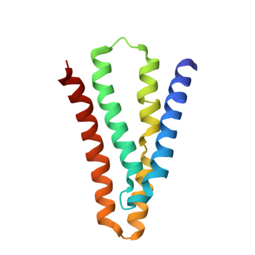
Molecular determinants of permeation in a fluoride-specific ion channel.

Last, N.B.Sun, S.Pham, M.C.Miller, C.

(2017) Elife 6

  • DOI: https://doi.org/10.7554/eLife.31259
  • Primary Citation of Related Structures:  
    6B24, 6B2A, 6B2B, 6B2D

  • PubMed Abstract: 

    Fluoride ion channels of the Fluc family combat toxicity arising from accumulation of environmental F - . Although crystal structures are known, the densely packed pore region has precluded delineation of the ion pathway. Here we chart out the Fluc pore and characterize its chemical requirements for transport. A ladder of H-bond donating residues creates a 'polar track' demarking the ion-conduction pathway. Surprisingly, while track polarity is well conserved, polarity is nonetheless functionally dispensable at several positions. A threonine at one end of the pore engages in vital interactions through its β-branched methyl group. Two critical central phenylalanines that directly coordinate F - through a quadrupolar-ion interaction cannot be functionally substituted by aromatic, non-polar, or polar sidechains. The only functional replacement is methionine, which coordinates F - through its partially positive γ-methylene in mimicry of phenylalanine's quadrupolar interaction. These results demonstrate the unusual chemical requirements for selectively transporting the strongly H-bonding F - anion.


  • Organizational Affiliation

    Department of Biochemistry, Howard Hughes Medical Institute, Brandeis University, Waltham, United States.


Macromolecules
Find similar proteins by:  (by identity cutoff)  |  3D Structure
Entity ID: 1
MoleculeChains Sequence LengthOrganismDetailsImage
Fluoride ion transporter CrcB
A, B
126Escherichia coliMutation(s): 2 
Gene Names: 
Membrane Entity: Yes 
UniProt
Find proteins for Q6J5N4 (Escherichia coli)
Explore Q6J5N4 
Go to UniProtKB:  Q6J5N4
Entity Groups  
Sequence Clusters30% Identity50% Identity70% Identity90% Identity95% Identity100% Identity
UniProt GroupQ6J5N4
Sequence Annotations
Expand
  • Reference Sequence
Find similar proteins by:  (by identity cutoff)  |  3D Structure
Entity ID: 2
MoleculeChains Sequence LengthOrganismDetailsImage
monobody
C, D
96Homo sapiensMutation(s): 0 
Entity Groups  
Sequence Clusters30% Identity50% Identity70% Identity90% Identity95% Identity100% Identity
Sequence Annotations
Expand
  • Reference Sequence
Experimental Data & Validation

Experimental Data

  • Method: X-RAY DIFFRACTION
  • Resolution: 3.01 Å
  • R-Value Free: 0.254 
  • R-Value Work: 0.238 
  • R-Value Observed: 0.238 
  • Space Group: P 41
Unit Cell:
Length ( Å )Angle ( ˚ )
a = 87.09α = 90
b = 87.09β = 90
c = 143.87γ = 90
Software Package:
Software NamePurpose
REFMACrefinement
Aimlessdata scaling
PDB_EXTRACTdata extraction
XDSdata reduction
PHASERphasing

Structure Validation

View Full Validation Report


Ligand Structure Quality Assessment 


Entry History & Funding Information

Deposition Data


Funding OrganizationLocationGrant Number
Howard Hughes Medical Institute (HHMI)United States--
National Institutes of Health/National Institute of General Medical Sciences (NIH/NIGMS)United StatesGM107023

Revision History  (Full details and data files)

  • Version 1.0: 2017-10-11
    Type: Initial release
  • Version 1.1: 2019-11-20
    Changes: Author supporting evidence
  • Version 1.2: 2023-10-04
    Changes: Data collection, Database references, Derived calculations, Refinement description