Molecular basis for feedback inhibition of tyrosine-regulated 3-deoxy-d-arabino-heptulosonate-7-phosphate synthase from Escherichia coli.
Cui, D., Deng, A., Bai, H., Yang, Z., Liang, Y., Liu, Z., Qiu, Q., Wang, L., Liu, S., Zhang, Y., Shi, Y., Qi, J., Wen, T.(2019) J Struct Biol 206: 322-334
- PubMed: 30946901
- DOI: https://doi.org/10.1016/j.jsb.2019.04.001
- Primary Citation of Related Structures:
6AGL, 6AGM - PubMed Abstract:
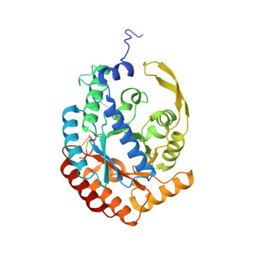
3-Deoxy-d-arabino-heptulosonate-7-phosphate synthase (DAHPS) is responsible for the biosynthesis of essential aromatic compounds in microorganisms and plants. It plays a crucial role in the regulation of the carbon flow into the shikimate pathway. Until now, the crystal structures and regulatory mechanisms of dimeric DAHPS enzymes from type Iα subclass have not been reported. Here, we reported dimeric structures of the tyrosine-regulated DAHPS from Escherichia coli, both in its apo form and complex with the inhibitor tyrosine at 2.5 and 2.0 Å resolutions, respectively. DAHPS(Tyr) has a typical (β/α) 8 TIM barrel, which is decorated with an N-terminal extension and an antiparallel β sheet, β6a/β6b. Inhibitor tyrosine binds at a cavity formed by residues of helices α3, α4, strands β6a, β6b and the adjacent loops, and directly interacts with residues P148, Q152, S181, I213 and N8 * . Although the small angle X-ray scattering profiles from DAHPS(Tyr) with and without tyrosine shows that tyrosine binding leaves most of DAHPS(Tyr) structures unaffected. The comparison of the liganded and unliganded crystal structures reveals that conformational changes of residues P148, Q152 and I213 initiate a transmission pathway to propagate the allosteric signal from the tyrosine-binding site to the active site, which is different from DAHPS(Phe), a phenylalanine-regulated isozyme from E. coli. In addition, mutations of five tyrosine-binding residues P148, Q152, S181, I213 and N8 * leads to tyrosine-resistant DAHPS(Tyr) enzymes. These findings provide a new insight into the regulatory mechanism of DAHPS enzymes and a basis for further engineering studies.
Organizational Affiliation:
CAS Key Laboratory of Pathogenic Microbiology and Immunology, Institute of Microbiology, Chinese Academy of Sciences, Beijing 100101, China; University of Chinese Academy of Sciences, Beijing 100049, China.