


Structural and Functional characterization of As1 from Plasmodium falciparum
Srivastava, D.K., Gunjan, S., Seshadri, V., Roy, S.To be published.
Experimental Data Snapshot
wwPDB Validation 3D Report Full Report

Entity ID: 1 | |||||
|---|---|---|---|---|---|
| Molecule | Chains | Sequence Length | Organism | Details | Image |
| Histone chaperone ASF1, putative | 161 | Plasmodium falciparum 3D7 | Mutation(s): 0 Gene Names: PF3D7_1224500 | 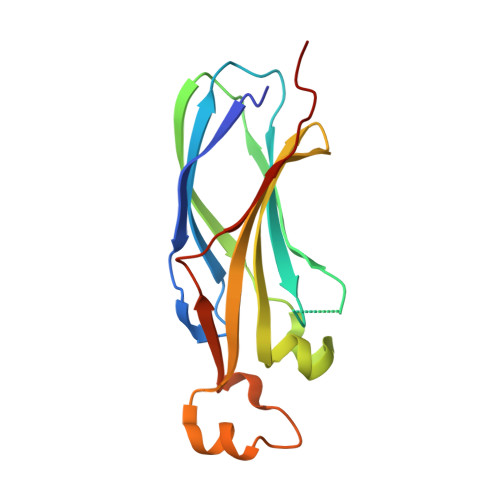 | |
UniProt | |||||
Find proteins for Q8I5H2 (Plasmodium falciparum (isolate 3D7)) Explore Q8I5H2 Go to UniProtKB: Q8I5H2 | |||||
Entity Groups | |||||
| Sequence Clusters | 30% Identity50% Identity70% Identity90% Identity95% Identity100% Identity | ||||
| UniProt Group | Q8I5H2 | ||||
Sequence AnnotationsExpand | |||||
| |||||
| Ligands 2 Unique | |||||
|---|---|---|---|---|---|
| ID | Chains | Name / Formula / InChI Key | 2D Diagram | 3D Interactions | |
| EDO Query on EDO | E [auth A], I [auth B] | 1,2-ETHANEDIOL C2 H6 O2 LYCAIKOWRPUZTN-UHFFFAOYSA-N |  | ||
| SCN Query on SCN | C [auth A] D [auth A] F [auth A] G [auth B] H [auth B] | THIOCYANATE ION C N S ZMZDMBWJUHKJPS-UHFFFAOYSA-M |  | ||
| Length ( Å ) | Angle ( ˚ ) |
|---|---|
| a = 62.733 | α = 90 |
| b = 62.733 | β = 90 |
| c = 98.725 | γ = 90 |
| Software Name | Purpose |
|---|---|
| PHENIX | refinement |
| XDS | data reduction |
| Aimless | data scaling |
| MrBUMP | phasing |
RCSB PDB (citation) is hosted by
RCSB PDB is a member of the

