6A2S

Mycobacterium tuberculosis LexA C-domain S160A


Experimental Data Snapshot

  • Method: X-RAY DIFFRACTION
  • Resolution: 2.50 Å
  • R-Value Free: 0.290 
  • R-Value Work: 0.241 
  • R-Value Observed: 0.244 

wwPDB Validation   3D Report Full Report


This is version 1.2 of the entry. See complete history


Literature

Biochemical characterization of Mycobacterium tuberculosis LexA and structural studies of its C-terminal segment.

Chandran, A.V.Srikalaivani, R.Paul, A.Vijayan, M.

(2019) Acta Crystallogr D Struct Biol 75: 41-55

  • DOI: https://doi.org/10.1107/S2059798318016066
  • Primary Citation of Related Structures:  
    6A2Q, 6A2R, 6A2S, 6A2T

  • PubMed Abstract: 

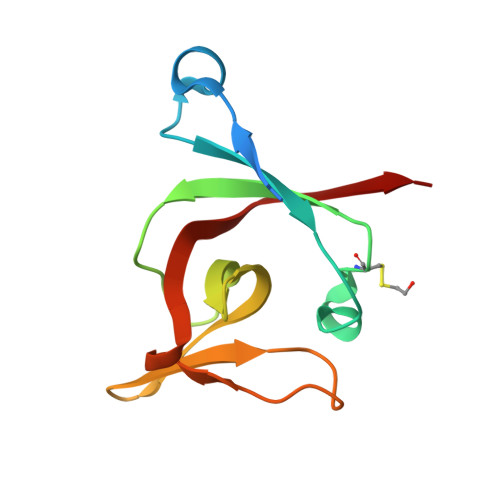
    LexA is a protein that is involved in the SOS response. The protein from Mycobacterium tuberculosis and its mutants have been biochemically characterized and the structures of their catalytic segments have been determined. The protein is made up of an N-terminal segment, which includes the DNA-binding domain, and a C-terminal segment encompassing much of the catalytic domain. The two segments are defined by a cleavage site. Full-length LexA, the two segments, two point mutants involving changes in the active-site residues (S160A and K197A) and another mutant involving a change at the cleavage site (G126D) were cloned and purified. The wild-type protein autocleaves at basic pH, while the mutants do not. The wild-type and the mutant proteins dimerize and bind DNA with equal facility. The C-terminal segment also dimerizes, and it also shows a tendency to form tetramers. The C-terminal segment readily crystallized. The crystals obtained from attempts involving the full-length protein and its mutants contained only the C-terminal segment including the catalytic core and a few residues preceding it, in a dimeric or tetrameric form, indicating protein cleavage during the long period involved in crystal formation. Modes of tetramerization of the full-length protein similar to those observed for the catalytic core are feasible. A complex of M. tuberculosis LexA and the cognate SOS box could be modeled in which the mutual orientation of the two N-terminal domains differs from that in the Escherichia coli LexA-DNA complex. These results represent the first thorough characterization of M. tuberculosis LexA and provide definitive information on its structure and assembly. They also provide leads for further exploration of this important protein.


  • Organizational Affiliation

    Molecular Biophysics Unit, Indian Institute of Science, Bangalore 560 012, India.


Macromolecules
Find similar proteins by:  (by identity cutoff)  |  3D Structure
Entity ID: 1
MoleculeChains Sequence LengthOrganismDetailsImage
LexA repressor
A, B, C, D, E
A, B, C, D, E, F, G, H
111Mycobacterium tuberculosis H37RvMutation(s): 1 
Gene Names: lexARv2720MTCY05A6.41
EC: 3.4.21.88
UniProt
Find proteins for P9WHR7 (Mycobacterium tuberculosis (strain ATCC 25618 / H37Rv))
Explore P9WHR7 
Go to UniProtKB:  P9WHR7
Entity Groups  
Sequence Clusters30% Identity50% Identity70% Identity90% Identity95% Identity100% Identity
UniProt GroupP9WHR7
Sequence Annotations
Expand
  • Reference Sequence
Small Molecules
Ligands 2 Unique
IDChains Name / Formula / InChI Key2D Diagram3D Interactions
P6G
Query on P6G

Download Ideal Coordinates CCD File 
K [auth A],
O [auth C]
HEXAETHYLENE GLYCOL
C12 H26 O7
IIRDTKBZINWQAW-UHFFFAOYSA-N
PEG
Query on PEG

Download Ideal Coordinates CCD File 
I [auth A]
J [auth A]
L [auth B]
M [auth B]
N [auth C]
I [auth A],
J [auth A],
L [auth B],
M [auth B],
N [auth C],
P [auth D],
Q [auth E],
R [auth F]
DI(HYDROXYETHYL)ETHER
C4 H10 O3
MTHSVFCYNBDYFN-UHFFFAOYSA-N
Modified Residues  1 Unique
IDChains TypeFormula2D DiagramParent
CME
Query on CME
A, B, C, D, E
A, B, C, D, E, F, G, H
L-PEPTIDE LINKINGC5 H11 N O3 S2CYS
Experimental Data & Validation

Experimental Data

  • Method: X-RAY DIFFRACTION
  • Resolution: 2.50 Å
  • R-Value Free: 0.290 
  • R-Value Work: 0.241 
  • R-Value Observed: 0.244 
  • Space Group: P 1 21 1
Unit Cell:
Length ( Å )Angle ( ˚ )
a = 57.61α = 90
b = 104.2β = 104.08
c = 88.1γ = 90
Software Package:
Software NamePurpose
REFMACrefinement
MOSFLMdata reduction
SCALAdata scaling
PHASERphasing

Structure Validation

View Full Validation Report


Entry History 

Deposition Data

Revision History  (Full details and data files)

  • Version 1.0: 2019-01-23
    Type: Initial release
  • Version 1.1: 2019-11-20
    Changes: Database references
  • Version 1.2: 2024-04-03
    Changes: Data collection, Database references, Refinement description