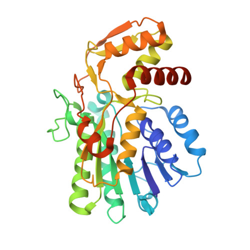
Crystallographic snapshots of UDP-glucuronic acid 4-epimerase ligand binding, rotation, and reduction.
Iacovino, L.G., Savino, S., Borg, A.J.E., Binda, C., Nidetzky, B., Mattevi, A.(2020) J Biol Chem 295: 12461-12473
- PubMed: 32661196
- DOI: https://doi.org/10.1074/jbc.RA120.014692
- Primary Citation of Related Structures:
6ZL6, 6ZLA, 6ZLD, 6ZLJ, 6ZLK, 6ZLL - PubMed Abstract:
UDP-glucuronic acid is converted to UDP-galacturonic acid en route to a variety of sugar-containing metabolites. This reaction is performed by a NAD + -dependent epimerase belonging to the short-chain dehydrogenase/reductase family. We present several high-resolution crystal structures of the UDP-glucuronic acid epimerase from Bacillus cereus The geometry of the substrate-NAD + interactions is finely arranged to promote hydride transfer. The exquisite complementarity between glucuronic acid and its binding site is highlighted by the observation that the unligated cavity is occupied by a cluster of ordered waters whose positions overlap the polar groups of the sugar substrate. Co-crystallization experiments led to a structure where substrate- and product-bound enzymes coexist within the same crystal. This equilibrium structure reveals the basis for a "swing and flip" rotation of the pro-chiral 4-keto-hexose-uronic acid intermediate that results from glucuronic acid oxidation, placing the C4' atom in position for receiving a hydride ion on the opposite side of the sugar ring. The product-bound active site is almost identical to that of the substrate-bound structure and satisfies all hydrogen-bonding requirements of the ligand. The structure of the apoenzyme together with the kinetic isotope effect and mutagenesis experiments further outlines a few flexible loops that exist in discrete conformations, imparting structural malleability required for ligand rotation while avoiding leakage of the catalytic intermediate and/or side reactions. These data highlight the double nature of the enzymatic mechanism: the active site features a high degree of precision in substrate recognition combined with the flexibility required for intermediate rotation.
Organizational Affiliation:
Department of Biology and Biotechnology "Lazzaro Spallanzani", University of Pavia, Pavia, Italy.


