6YU4

Crystal structure of MhsT in complex with L-4F-phenylalanine


Experimental Data Snapshot

  • Method: X-RAY DIFFRACTION
  • Resolution: 2.26 Å
  • R-Value Free: 0.261 
  • R-Value Work: 0.223 
  • R-Value Observed: 0.225 

wwPDB Validation   3D Report Full Report


Ligand Structure Quality Assessment 


This is version 1.3 of the entry. See complete history


Literature

A non-helical region in transmembrane helix 6 of hydrophobic amino acid transporter MhsT mediates substrate recognition.

Focht, D.Neumann, C.Lyons, J.Eguskiza Bilbao, A.Blunck, R.Malinauskaite, L.Schwarz, I.O.Javitch, J.A.Quick, M.Nissen, P.

(2021) EMBO J 40: e105164-e105164

  • DOI: https://doi.org/10.15252/embj.2020105164
  • Primary Citation of Related Structures:  
    6YU2, 6YU3, 6YU4, 6YU5, 6YU6, 6YU7

  • PubMed Abstract: 

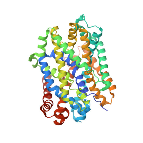
    MhsT of Bacillus halodurans is a transporter of hydrophobic amino acids and a homologue of the eukaryotic SLC6 family of Na + -dependent symporters for amino acids, neurotransmitters, osmolytes, or creatine. The broad range of transported amino acids by MhsT prompted the investigation of the substrate recognition mechanism. Here, we report six new substrate-bound structures of MhsT, which, in conjunction with functional studies, reveal how the flexibility of a Gly-Met-Gly (GMG) motif in the unwound region of transmembrane segment 6 (TM6) is central for the recognition of substrates of different size by tailoring the binding site shape and volume. MhsT mutants, harboring substitutions within the unwound GMG loop and substrate binding pocket that mimick the binding sites of eukaryotic SLC6A18/B0AT3 and SLC6A19/B0AT1 transporters of neutral amino acids, exhibited impaired transport of aromatic amino acids that require a large binding site volume. Conservation of a general (G/A/C)ΦG motif among eukaryotic members of SLC6 family suggests a role for this loop in a common mechanism for substrate recognition and translocation by SLC6 transporters of broad substrate specificity.


  • Organizational Affiliation

    Department of Molecular Biology and Genetics, Danish Research Institute of Translational Neuroscience-DANDRITE, Nordic-EMBL Partnership for Molecular Medicine, Aarhus University, Aarhus C, Denmark.


Macromolecules
Find similar proteins by:  (by identity cutoff)  |  3D Structure
Entity ID: 1
MoleculeChains Sequence LengthOrganismDetailsImage
Sodium-dependent transporter455Halalkalibacterium haloduransMutation(s): 0 
Gene Names: E2L07_01100
Membrane Entity: Yes 
UniProt
Find proteins for Q9KDT3 (Halalkalibacterium halodurans (strain ATCC BAA-125 / DSM 18197 / FERM 7344 / JCM 9153 / C-125))
Explore Q9KDT3 
Go to UniProtKB:  Q9KDT3
Entity Groups  
Sequence Clusters30% Identity50% Identity70% Identity90% Identity95% Identity100% Identity
UniProt GroupQ9KDT3
Sequence Annotations
Expand
  • Reference Sequence
Small Molecules
Ligands 5 Unique
IDChains Name / Formula / InChI Key2D Diagram3D Interactions
PCW
Query on PCW

Download Ideal Coordinates CCD File 
E [auth A]1,2-DIOLEOYL-SN-GLYCERO-3-PHOSPHOCHOLINE
C44 H85 N O8 P
SNKAWJBJQDLSFF-NVKMUCNASA-O
LMT
Query on LMT

Download Ideal Coordinates CCD File 
F [auth A]
G [auth A]
H [auth A]
I [auth A]
L [auth A]
F [auth A],
G [auth A],
H [auth A],
I [auth A],
L [auth A],
M [auth A]
DODECYL-BETA-D-MALTOSIDE
C24 H46 O11
NLEBIOOXCVAHBD-QKMCSOCLSA-N
SOG
Query on SOG

Download Ideal Coordinates CCD File 
J [auth A],
K [auth A]
octyl 1-thio-beta-D-glucopyranoside
C14 H28 O5 S
CGVLVOOFCGWBCS-RGDJUOJXSA-N
PFF (Subject of Investigation/LOI)
Query on PFF

Download Ideal Coordinates CCD File 
D [auth A]4-FLUORO-L-PHENYLALANINE
C9 H10 F N O2
XWHHYOYVRVGJJY-QMMMGPOBSA-N
NA (Subject of Investigation/LOI)
Query on NA

Download Ideal Coordinates CCD File 
B [auth A],
C [auth A]
SODIUM ION
Na
FKNQFGJONOIPTF-UHFFFAOYSA-N
Experimental Data & Validation

Experimental Data

  • Method: X-RAY DIFFRACTION
  • Resolution: 2.26 Å
  • R-Value Free: 0.261 
  • R-Value Work: 0.223 
  • R-Value Observed: 0.225 
  • Space Group: P 1 2 1
Unit Cell:
Length ( Å )Angle ( ˚ )
a = 44.15α = 90
b = 49.85β = 96.08
c = 109.73γ = 90
Software Package:
Software NamePurpose
PHENIXrefinement
PDB_EXTRACTdata extraction
XDSdata reduction
XSCALEdata scaling
PHENIXphasing

Structure Validation

View Full Validation Report


Ligand Structure Quality Assessment 


Entry History & Funding Information

Deposition Data


Funding OrganizationLocationGrant Number
LundbeckfondenDenmark2011-3868
LundbeckfondenDenmark2015-2704
LundbeckfondenDenmark2015-3225
LundbeckfondenDenmark2016-2518

Revision History  (Full details and data files)

  • Version 1.0: 2020-07-15
    Type: Initial release
  • Version 1.1: 2020-07-29
    Type: Remediation
    Reason: Carbohydrate remediation
    Changes: Derived calculations, Structure summary
  • Version 1.2: 2021-01-27
    Changes: Database references, Structure summary
  • Version 1.3: 2024-01-24
    Changes: Data collection, Database references, Refinement description