6YC4

Crystal structure of the steady-state activated state of the light-driven sodium pump KR2 in the pentameric form at room temperature, pH 8.0


Experimental Data Snapshot

  • Method: X-RAY DIFFRACTION
  • Resolution: 2.60 Å
  • R-Value Free: 0.212 
  • R-Value Work: 0.185 
  • R-Value Observed: 0.186 

wwPDB Validation   3D Report Full Report


This is version 1.2 of the entry. See complete history


Literature

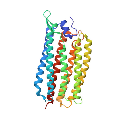
Molecular mechanism of light-driven sodium pumping.

Kovalev, K.Astashkin, R.Gushchin, I.Orekhov, P.Volkov, D.Zinovev, E.Marin, E.Rulev, M.Alekseev, A.Royant, A.Carpentier, P.Vaganova, S.Zabelskii, D.Baeken, C.Sergeev, I.Balandin, T.Bourenkov, G.Carpena, X.Boer, R.Maliar, N.Borshchevskiy, V.Buldt, G.Bamberg, E.Gordeliy, V.

(2020) Nat Commun 11: 2137-2137

  • DOI: https://doi.org/10.1038/s41467-020-16032-y
  • Primary Citation of Related Structures:  
    6YBY, 6YC0, 6YC2, 6YC3, 6YC4

  • PubMed Abstract: 

    The light-driven sodium-pumping rhodopsin KR2 from Krokinobacter eikastus is the only non-proton cation active transporter with demonstrated potential for optogenetics. However, the existing structural data on KR2 correspond exclusively to its ground state, and show no sodium inside the protein, which hampers the understanding of sodium-pumping mechanism. Here we present crystal structure of the O-intermediate of the physiologically relevant pentameric form of KR2 at the resolution of 2.1 Å, revealing a sodium ion near the retinal Schiff base, coordinated by N112 and D116 of the characteristic NDQ triad. We also obtained crystal structures of D116N and H30A variants, conducted metadynamics simulations and measured pumping activities of putative pathway mutants to demonstrate that sodium release likely proceeds alongside Q78 towards the structural sodium ion bound between KR2 protomers. Our findings highlight the importance of pentameric assembly for sodium pump function, and may be used for rational engineering of enhanced optogenetic tools.


  • Organizational Affiliation

    Institut de Biologie Structurale (IBS), Université Grenoble Alpes, CEA, CNRS, Grenoble, France.


Macromolecules
Find similar proteins by:  (by identity cutoff)  |  3D Structure
Entity ID: 1
MoleculeChains Sequence LengthOrganismDetailsImage
Sodium pumping rhodopsin
A, B, C, D, E
273Dokdonia eikastaMutation(s): 0 
Gene Names: NaR
Membrane Entity: Yes 
UniProt
Find proteins for N0DKS8 (Dokdonia eikasta)
Explore N0DKS8 
Go to UniProtKB:  N0DKS8
Entity Groups  
Sequence Clusters30% Identity50% Identity70% Identity90% Identity95% Identity100% Identity
UniProt GroupN0DKS8
Sequence Annotations
Expand
  • Reference Sequence
Small Molecules
Ligands 6 Unique
IDChains Name / Formula / InChI Key2D Diagram3D Interactions
OLC
Query on OLC

Download Ideal Coordinates CCD File 
AB [auth D]
BB [auth D]
F [auth A]
FA [auth B]
G [auth A]
AB [auth D],
BB [auth D],
F [auth A],
FA [auth B],
G [auth A],
H [auth A],
I [auth A],
J [auth A],
JB [auth D],
KA [auth C],
LA [auth C],
MA [auth C],
MB [auth E],
NA [auth C],
OA [auth C],
QB [auth E],
R [auth A],
RB [auth E],
SB [auth E],
T [auth A],
TB [auth E],
W [auth B],
WA [auth C],
X [auth B],
Y [auth B],
YA [auth D],
Z [auth B],
ZA [auth D]
(2R)-2,3-dihydroxypropyl (9Z)-octadec-9-enoate
C21 H40 O4
RZRNAYUHWVFMIP-GDCKJWNLSA-N
RET
Query on RET

Download Ideal Coordinates CCD File 
AC [auth E],
EA [auth B],
IB [auth D],
S [auth A],
VA [auth C]
RETINAL
C20 H28 O
NCYCYZXNIZJOKI-OVSJKPMPSA-N
LFA
Query on LFA

Download Ideal Coordinates CCD File 
AA [auth B]
BA [auth B]
CA [auth B]
CB [auth D]
DB [auth D]
AA [auth B],
BA [auth B],
CA [auth B],
CB [auth D],
DB [auth D],
EB [auth D],
FB [auth D],
GB [auth D],
K [auth A],
L [auth A],
M [auth A],
N [auth A],
O [auth A],
P [auth A],
PA [auth C],
QA [auth C],
RA [auth C],
UB [auth E],
VB [auth E],
WB [auth E],
XB [auth E]
EICOSANE
C20 H42
CBFCDTFDPHXCNY-UHFFFAOYSA-N
OLA
Query on OLA

Download Ideal Coordinates CCD File 
GA [auth B]
HA [auth B]
KB [auth D]
NB [auth E]
OB [auth E]
GA [auth B],
HA [auth B],
KB [auth D],
NB [auth E],
OB [auth E],
PB [auth E],
TA [auth C],
ZB [auth E]
OLEIC ACID
C18 H34 O2
ZQPPMHVWECSIRJ-KTKRTIGZSA-N
GOL
Query on GOL

Download Ideal Coordinates CCD File 
IA [auth B],
U [auth A],
UA [auth C]
GLYCEROL
C3 H8 O3
PEDCQBHIVMGVHV-UHFFFAOYSA-N
NA (Subject of Investigation/LOI)
Query on NA

Download Ideal Coordinates CCD File 
BC [auth E]
DA [auth B]
HB [auth D]
JA [auth B]
LB [auth D]
BC [auth E],
DA [auth B],
HB [auth D],
JA [auth B],
LB [auth D],
Q [auth A],
SA [auth C],
V [auth A],
XA [auth C],
YB [auth E]
SODIUM ION
Na
FKNQFGJONOIPTF-UHFFFAOYSA-N
Experimental Data & Validation

Experimental Data

  • Method: X-RAY DIFFRACTION
  • Resolution: 2.60 Å
  • R-Value Free: 0.212 
  • R-Value Work: 0.185 
  • R-Value Observed: 0.186 
  • Space Group: C 2 2 21
Unit Cell:
Length ( Å )Angle ( ˚ )
a = 135.189α = 90
b = 239.861β = 90
c = 138.456γ = 90
Software Package:
Software NamePurpose
Aimlessdata scaling
REFMACrefinement
PDB_EXTRACTdata extraction
XDSdata reduction
MOLREPphasing

Structure Validation

View Full Validation Report


Entry History & Funding Information

Deposition Data


Funding OrganizationLocationGrant Number
French National Research AgencyFranceANR-15-CE11-0029-02
French National Research AgencyFranceANR-10-INBS-05-02
Grenoble Alliance for Integrated Structural Cell Biology (GRAL)FranceANR-17-EURE-0003
Russian Science FoundationRussian Federation16-15-00242
Russian Science FoundationRussian Federation6.3157.2017/PP
Russian Foundation for Basic ResearchRussian Federation19-29-12022

Revision History  (Full details and data files)

  • Version 1.0: 2020-04-08
    Type: Initial release
  • Version 1.1: 2020-05-13
    Changes: Database references
  • Version 1.2: 2024-01-24
    Changes: Data collection, Database references, Derived calculations, Refinement description