6XX7

Arabidopsis thaliana Casein Kinase 2 (CK2) alpha-1 crystal in complex with ANP


Experimental Data Snapshot

  • Method: X-RAY DIFFRACTION
  • Resolution: 2.40 Å
  • R-Value Free: 0.246 
  • R-Value Work: 0.195 
  • R-Value Observed: 0.198 

wwPDB Validation   3D Report Full Report


Ligand Structure Quality Assessment 


This is version 1.1 of the entry. See complete history


Literature

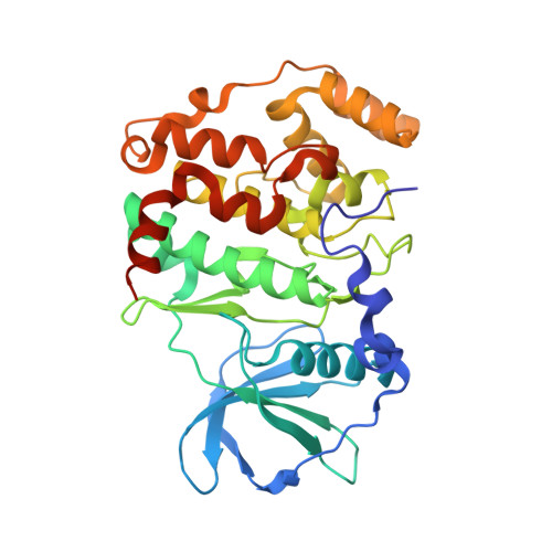
Crystal structure of Arabidopsis thaliana casein kinase 2 alpha 1.

Demulder, M.De Veylder, L.Loris, R.

(2020) Acta Crystallogr F Struct Biol Commun 76: 182-191

  • DOI: https://doi.org/10.1107/S2053230X20004537
  • Primary Citation of Related Structures:  
    6XX6, 6XX7, 6XX8, 6XX9

  • PubMed Abstract: 

    Casein kinase 2 (CK2) is a ubiquitous pleiotropic enzyme that is highly conserved across eukaryotic kingdoms. CK2 is singular amongst kinases as it is highly rigid and constitutively active. Arabidopsis thaliana is widely used as a model system in molecular plant research; the biological functions of A. thaliana CK2 are well studied in vivo and many of its substrates have been identified. Here, crystal structures of the α subunit of A. thaliana CK2 in three crystal forms and of its complex with the nonhydrolyzable ATP analog AMppNHp are presented. While the C-lobe of the enzyme is highly rigid, structural plasticity is observed for the N-lobe. Small but significant displacements within the active cleft are necessary in order to avoid steric clashes with the AMppNHp molecule. Binding of AMppNHp is influenced by a rigid-body motion of the N-lobe that was not previously recognized in maize CK2.


  • Organizational Affiliation

    Structural Biology Brussels, Vrije Universiteit Brussel, Pleinlaan 2, B-1050 Brussels, Belgium.


Macromolecules
Find similar proteins by:  (by identity cutoff)  |  3D Structure
Entity ID: 1
MoleculeChains Sequence LengthOrganismDetailsImage
Casein kinase II subunit alpha-1342Arabidopsis thalianaMutation(s): 0 
Gene Names: CKA1At5g67380K8K14.10
EC: 2.7.11.1
UniProt
Find proteins for Q08467 (Arabidopsis thaliana)
Explore Q08467 
Go to UniProtKB:  Q08467
Entity Groups  
Sequence Clusters30% Identity50% Identity70% Identity90% Identity95% Identity100% Identity
UniProt GroupQ08467
Sequence Annotations
Expand
  • Reference Sequence
Small Molecules
Ligands 5 Unique
IDChains Name / Formula / InChI Key2D Diagram3D Interactions
ANP (Subject of Investigation/LOI)
Query on ANP

Download Ideal Coordinates CCD File 
B [auth A]PHOSPHOAMINOPHOSPHONIC ACID-ADENYLATE ESTER
C10 H17 N6 O12 P3
PVKSNHVPLWYQGJ-KQYNXXCUSA-N
PGE
Query on PGE

Download Ideal Coordinates CCD File 
H [auth A]TRIETHYLENE GLYCOL
C6 H14 O4
ZIBGPFATKBEMQZ-UHFFFAOYSA-N
SO4
Query on SO4

Download Ideal Coordinates CCD File 
D [auth A]SULFATE ION
O4 S
QAOWNCQODCNURD-UHFFFAOYSA-L
CL
Query on CL

Download Ideal Coordinates CCD File 
E [auth A],
F [auth A],
G [auth A]
CHLORIDE ION
Cl
VEXZGXHMUGYJMC-UHFFFAOYSA-M
MG
Query on MG

Download Ideal Coordinates CCD File 
C [auth A]MAGNESIUM ION
Mg
JLVVSXFLKOJNIY-UHFFFAOYSA-N
Experimental Data & Validation

Experimental Data

  • Method: X-RAY DIFFRACTION
  • Resolution: 2.40 Å
  • R-Value Free: 0.246 
  • R-Value Work: 0.195 
  • R-Value Observed: 0.198 
  • Space Group: P 41 21 2
Unit Cell:
Length ( Å )Angle ( ˚ )
a = 60.56α = 90
b = 60.56β = 90
c = 228.6γ = 90
Software Package:
Software NamePurpose
PHASERphasing
PHENIXrefinement
XDSdata reduction
XDSdata scaling

Structure Validation

View Full Validation Report


Ligand Structure Quality Assessment 


Entry History & Funding Information

Deposition Data


Funding OrganizationLocationGrant Number
Research Foundation - Flanders (FWO)BelgiumG023616N
Research Foundation - Flanders (FWO)BelgiumG011420N

Revision History  (Full details and data files)

  • Version 1.0: 2020-04-15
    Type: Initial release
  • Version 1.1: 2024-01-24
    Changes: Data collection, Database references, Refinement description