6X6O

Crystal structure of T4 protein Spackle as determined by native SAD phasing


Experimental Data Snapshot

  • Method: X-RAY DIFFRACTION
  • Resolution: 1.52 Å
  • R-Value Free: 0.191 
  • R-Value Work: 0.173 
  • R-Value Observed: 0.174 

wwPDB Validation   3D Report Full Report


This is version 1.0 of the entry. See complete history


Literature

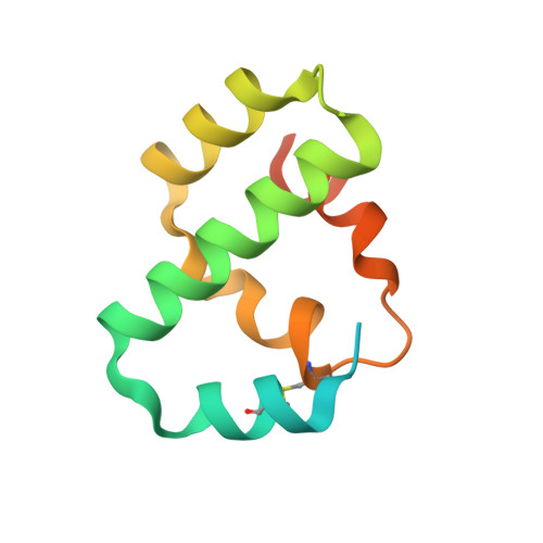
Crystal structure of bacteriophage T4 Spackle as determined by native SAD phasing.

Shi, K.Kurniawan, F.Banerjee, S.Moeller, N.H.Aihara, H.

(2020) Acta Crystallogr D Struct Biol 76: 899-904

  • DOI: https://doi.org/10.1107/S2059798320010979
  • Primary Citation of Related Structures:  
    6X6O

  • PubMed Abstract: 

    The crystal structure of a bacteriophage T4 early gene product, Spackle, was determined by native sulfur single-wavelength anomalous diffraction (SAD) phasing using synchrotron radiation and was refined to 1.52 Å resolution. The structure shows that Spackle consists of a bundle of five α-helices, forming a relatively flat disc-like overall shape. Although Spackle forms a dimer in the crystal, size-exclusion chromatography with multi-angle light scattering shows that it is monomeric in solution. Mass spectrometry confirms that purified mature Spackle lacks the amino-terminal signal peptide and contains an intramolecular disulfide bond, consistent with its proposed role in the periplasm of T4 phage-infected Escherichia coli cells. The surface electrostatic potential of Spackle shows a strikingly bipolar charge distribution, suggesting a possible mode of membrane association and inhibition of the tail lysozyme activity in T4 bacteriophage superinfection exclusion.


  • Organizational Affiliation

    Department of Biochemistry, Molecular Biology and Biophysics, University of Minnesota, Minneapolis, MN 55455, USA.


Macromolecules
Find similar proteins by:  (by identity cutoff)  |  3D Structure
Entity ID: 1
MoleculeChains Sequence LengthOrganismDetailsImage
Protein spackle
A, B
105Tequatrovirus T4Mutation(s): 0 
Gene Names: sp61.3
UniProt
Find proteins for P39230 (Enterobacteria phage T4)
Explore P39230 
Go to UniProtKB:  P39230
Entity Groups  
Sequence Clusters30% Identity50% Identity70% Identity90% Identity95% Identity100% Identity
UniProt GroupP39230
Sequence Annotations
Expand
  • Reference Sequence
Experimental Data & Validation

Experimental Data

  • Method: X-RAY DIFFRACTION
  • Resolution: 1.52 Å
  • R-Value Free: 0.191 
  • R-Value Work: 0.173 
  • R-Value Observed: 0.174 
  • Space Group: I 2 2 2
Unit Cell:
Length ( Å )Angle ( ˚ )
a = 44.424α = 90
b = 79.822β = 90
c = 93.227γ = 90
Software Package:
Software NamePurpose
PHENIXrefinement
XDSdata reduction
PDB_EXTRACTdata extraction
XDSdata scaling
SHELXDphasing

Structure Validation

View Full Validation Report


Entry History & Funding Information

Deposition Data


Funding OrganizationLocationGrant Number
National Institutes of Health/National Institute of General Medical Sciences (NIH/NIGMS)United StatesGM118047

Revision History  (Full details and data files)

  • Version 1.0: 2020-09-16
    Type: Initial release