6WO5

Structure of Hepatitis C Virus Envelope Glycoprotein E2 core from genotype 1a bound to neutralizing antibody 212.1.1 and non neutralizing antibody E1


Experimental Data Snapshot

  • Method: X-RAY DIFFRACTION
  • Resolution: 2.62 Å
  • R-Value Free: 0.250 
  • R-Value Work: 0.203 
  • R-Value Observed: 0.205 

wwPDB Validation   3D Report Full Report


Ligand Structure Quality Assessment 


This is version 1.1 of the entry. See complete history


Literature

An alternate conformation of HCV E2 neutralizing face as an additional vaccine target.

Tzarum, N.Giang, E.Kadam, R.U.Chen, F.Nagy, K.Augestad, E.H.Velazquez-Moctezuma, R.Keck, Z.Y.Hua, Y.Stanfield, R.L.Dreux, M.Prentoe, J.Foung, S.K.H.Bukh, J.Wilson, I.A.Law, M.

(2020) Sci Adv 6: eabb5642-eabb5642

  • DOI: https://doi.org/10.1126/sciadv.abb5642
  • Primary Citation of Related Structures:  
    6WO3, 6WO4, 6WO5, 6WOQ, 6WOR, 6WOS

  • PubMed Abstract: 

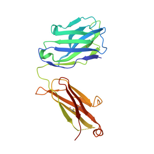
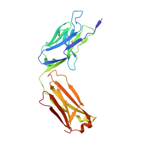
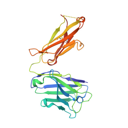
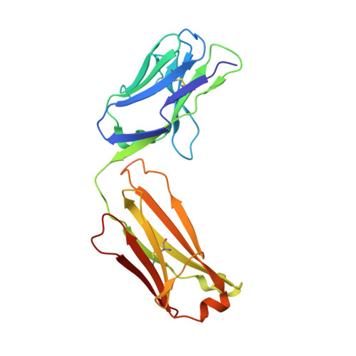
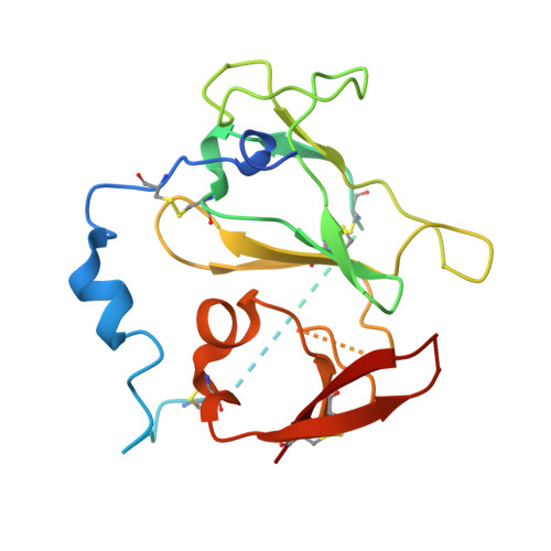
    To achieve global elimination of hepatitis C virus (HCV), an effective cross-genotype vaccine is needed. The HCV envelope glycoprotein E2 is the main target for neutralizing antibodies (nAbs), which aid in HCV clearance and protection. E2 is structurally flexible and functions in engaging host receptors. Many nAbs bind to the "neutralizing face" on E2, including several broadly nAbs encoded by the V H 1-69 germline gene family that bind to a similar conformation (A) of this face. Here, a previously unknown conformation (B) of the neutralizing face is revealed in crystal structures of two of four additional E2-V H 1-69 nAb complexes. In this conformation, the E2 front-layer region is displaced upon antibody binding, exposing residues in the back layer for direct antibody interaction. This E2 B structure may represent another conformational state in the viral entry process that is susceptible to antibody neutralization and thus provide a new target for rational vaccine development.


  • Organizational Affiliation

    Department of Integrative Structural and Computational Biology, The Scripps Research Institute, La Jolla, CA 92037, USA.


Macromolecules
Find similar proteins by:  (by identity cutoff)  |  3D Structure
Entity ID: 1
MoleculeChains Sequence LengthOrganismDetailsImage
Fab E1 heavy chain
A, C
227Homo sapiensMutation(s): 0 
Entity Groups  
Sequence Clusters30% Identity50% Identity70% Identity90% Identity95% Identity100% Identity
Sequence Annotations
Expand
  • Reference Sequence
Find similar proteins by:  (by identity cutoff)  |  3D Structure
Entity ID: 2
MoleculeChains Sequence LengthOrganismDetailsImage
Fab E1 light chain
B, D
221Homo sapiensMutation(s): 0 
Entity Groups  
Sequence Clusters30% Identity50% Identity70% Identity90% Identity95% Identity100% Identity
Sequence Annotations
Expand
  • Reference Sequence
Find similar proteins by:  (by identity cutoff)  |  3D Structure
Entity ID: 3
MoleculeChains Sequence LengthOrganismDetailsImage
Fab 212.1.1 heavy chainE [auth H],
G
230Homo sapiensMutation(s): 0 
Entity Groups  
Sequence Clusters30% Identity50% Identity70% Identity90% Identity95% Identity100% Identity
Sequence Annotations
Expand
  • Reference Sequence
Find similar proteins by:  (by identity cutoff)  |  3D Structure
Entity ID: 4
MoleculeChains Sequence LengthOrganismDetailsImage
Fab 212.1.1 light chainF [auth L],
H [auth I]
214Homo sapiensMutation(s): 0 
Entity Groups  
Sequence Clusters30% Identity50% Identity70% Identity90% Identity95% Identity100% Identity
Sequence Annotations
Expand
  • Reference Sequence
Find similar proteins by:  (by identity cutoff)  |  3D Structure
Entity ID: 5
MoleculeChains Sequence LengthOrganismDetailsImage
Envelope glycoprotein E2I [auth E],
J [auth F]
191Hepatitis C virus (isolate H)Mutation(s): 0 
UniProt
Find proteins for P27958 (Hepatitis C virus genotype 1a (isolate H77))
Explore P27958 
Go to UniProtKB:  P27958
Entity Groups  
Sequence Clusters30% Identity50% Identity70% Identity90% Identity95% Identity100% Identity
UniProt GroupP27958
Sequence Annotations
Expand
  • Reference Sequence
Oligosaccharides

Help

Entity ID: 6
MoleculeChains Length2D Diagram Glycosylation3D Interactions
beta-D-mannopyranose-(1-4)-2-acetamido-2-deoxy-beta-D-glucopyranose-(1-4)-2-acetamido-2-deoxy-beta-D-glucopyranoseK [auth J],
N
3N-Glycosylation
Glycosylation Resources
GlyTouCan:  G15407YE
GlyCosmos:  G15407YE
GlyGen:  G15407YE
Entity ID: 7
MoleculeChains Length2D Diagram Glycosylation3D Interactions
2-acetamido-2-deoxy-beta-D-glucopyranose-(1-4)-2-acetamido-2-deoxy-beta-D-glucopyranoseL [auth K],
M
2N-Glycosylation
Glycosylation Resources
GlyTouCan:  G42666HT
GlyCosmos:  G42666HT
GlyGen:  G42666HT
Entity ID: 8
MoleculeChains Length2D Diagram Glycosylation3D Interactions
alpha-D-mannopyranose-(1-3)-[alpha-D-mannopyranose-(1-6)]beta-D-mannopyranose-(1-4)-2-acetamido-2-deoxy-beta-D-glucopyranose-(1-4)-2-acetamido-2-deoxy-beta-D-glucopyranose
O
5N-Glycosylation
Glycosylation Resources
GlyTouCan:  G22768VO
GlyCosmos:  G22768VO
GlyGen:  G22768VO
Experimental Data & Validation

Experimental Data

  • Method: X-RAY DIFFRACTION
  • Resolution: 2.62 Å
  • R-Value Free: 0.250 
  • R-Value Work: 0.203 
  • R-Value Observed: 0.205 
  • Space Group: P 1 21 1
Unit Cell:
Length ( Å )Angle ( ˚ )
a = 55.793α = 90
b = 242.204β = 98.31
c = 100.729γ = 90
Software Package:
Software NamePurpose
PHENIXrefinement
HKL-2000data reduction
Blu-Icedata collection
SCALEPACKdata scaling
PHASERphasing

Structure Validation

View Full Validation Report


Ligand Structure Quality Assessment 


Entry History & Funding Information

Deposition Data


Funding OrganizationLocationGrant Number
National Institutes of Health/National Institute Of Allergy and Infectious Diseases (NIH/NIAID)United StatesAl123365
National Institutes of Health/National Institute Of Allergy and Infectious Diseases (NIH/NIAID)United StatesAl106005

Revision History  (Full details and data files)

  • Version 1.0: 2020-08-19
    Type: Initial release
  • Version 1.1: 2023-10-18
    Changes: Data collection, Database references, Refinement description