6WEY

High-resolution structure of the SARS-CoV-2 NSP3 Macro X domain


Experimental Data Snapshot

  • Method: X-RAY DIFFRACTION
  • Resolution: 0.95 Å
  • R-Value Free: 0.136 
  • R-Value Work: 0.119 
  • R-Value Observed: 0.120 

wwPDB Validation   3D Report Full Report


This is version 1.5 of the entry. See complete history


Literature

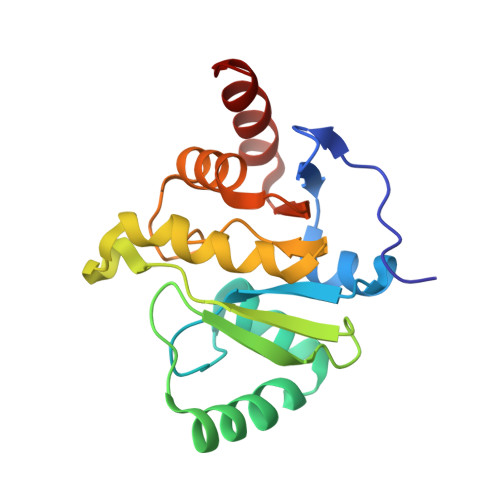
Molecular Basis for ADP-Ribose Binding to the Mac1 Domain of SARS-CoV-2 nsp3.

Frick, D.N.Virdi, R.S.Vuksanovic, N.Dahal, N.Silvaggi, N.R.

(2020) Biochemistry 59: 2608-2615

  • DOI: https://doi.org/10.1021/acs.biochem.0c00309
  • Primary Citation of Related Structures:  
    6WEY

  • PubMed Abstract: 

    The virus that causes COVID-19, SARS-CoV-2, has a large RNA genome that encodes numerous proteins that might be targets for antiviral drugs. Some of these proteins, such as the RNA-dependent RNA polymerase, helicase, and main protease, are well conserved between SARS-CoV-2 and the original SARS virus, but several others are not. This study examines one of the proteins encoded by SARS-CoV-2 that is most different, a macrodomain of nonstructural protein 3 (nsp3). Although 26% of the amino acids in this SARS-CoV-2 macrodomain differ from those observed in other coronaviruses, biochemical and structural data reveal that the protein retains the ability to bind ADP-ribose, which is an important characteristic of beta coronaviruses and a potential therapeutic target.


Macromolecules
Find similar proteins by:  (by identity cutoff)  |  3D Structure
Entity ID: 1
MoleculeChains Sequence LengthOrganismDetailsImage
Non-structural protein 3172Severe acute respiratory syndrome coronavirus 2Mutation(s): 0 
Gene Names: rep1a-1b
EC: 3.4.22
UniProt
Find proteins for P0DTD1 (Severe acute respiratory syndrome coronavirus 2)
Explore P0DTD1 
Go to UniProtKB:  P0DTD1
Entity Groups  
Sequence Clusters30% Identity50% Identity70% Identity90% Identity95% Identity100% Identity
UniProt GroupP0DTD1
Sequence Annotations
Expand
  • Reference Sequence
Experimental Data & Validation

Experimental Data

  • Method: X-RAY DIFFRACTION
  • Resolution: 0.95 Å
  • R-Value Free: 0.136 
  • R-Value Work: 0.119 
  • R-Value Observed: 0.120 
  • Space Group: P 21 21 21
Unit Cell:
Length ( Å )Angle ( ˚ )
a = 43.322α = 90
b = 54.424β = 90
c = 67.567γ = 90
Software Package:
Software NamePurpose
PHENIXrefinement
DIALSdata reduction
Aimlessdata scaling
PHASERphasing

Structure Validation

View Full Validation Report


Entry History & Funding Information

Deposition Data


Funding OrganizationLocationGrant Number
National Science Foundation (NSF, United States)United States1903899

Revision History  (Full details and data files)

  • Version 1.0: 2020-04-29
    Type: Initial release
  • Version 1.1: 2020-05-06
    Changes: Database references, Source and taxonomy, Structure summary
  • Version 1.2: 2020-08-26
    Changes: Database references
  • Version 1.3: 2020-09-02
    Changes: Source and taxonomy
  • Version 1.4: 2021-01-27
    Changes: Structure summary
  • Version 1.5: 2023-10-18
    Changes: Data collection, Database references, Refinement description