6W3Q

APE1 exonuclease substrate complex L104R


Experimental Data Snapshot

  • Method: X-RAY DIFFRACTION
  • Resolution: 2.49 Å
  • R-Value Free: 0.259 
  • R-Value Work: 0.222 
  • R-Value Observed: 0.224 

wwPDB Validation   3D Report Full Report


This is version 1.1 of the entry. See complete history


Literature

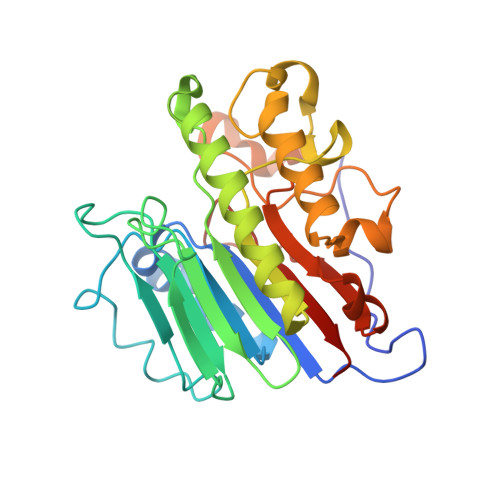
Molecular and structural characterization of disease-associated APE1 polymorphisms.

Whitaker, A.M.Stark, W.J.Flynn, T.S.Freudenthal, B.D.

(2020) DNA Repair (Amst) 91-92: 102867-102867

  • DOI: https://doi.org/10.1016/j.dnarep.2020.102867
  • Primary Citation of Related Structures:  
    6W0Q, 6W2P, 6W3L, 6W3N, 6W3Q, 6W3U, 6W43

  • PubMed Abstract: 

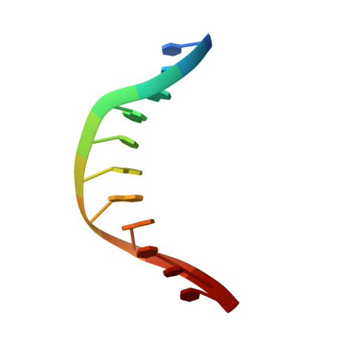
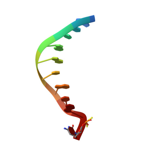
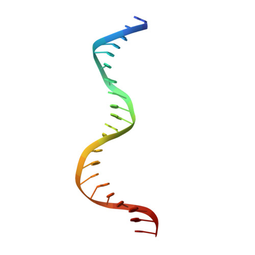
    Under conditions of oxidative stress, reactive oxygen species (ROS) continuously assault the structure of DNA resulting in oxidation and fragmentation of the nucleobases. When the nucleobase structure is altered, its base-pairing properties may also be altered, promoting mutations. Consequently, oxidative DNA damage is a major source of the mutation load that gives rise to numerous human maladies, including cancer. Base excision repair (BER) is the primary pathway tasked with removing and replacing mutagenic DNA base damage. Apurinic/apyrimidinic endonuclease 1 (APE1) is a central enzyme with AP-endonuclease and 3' to 5' exonuclease functions during BER, and therefore is key to maintenance of genome stability. Polymorphisms, or SNPs, in the gene encoding APE1 (APEX1) have been identified among specific human populations and result in variants of APE1 with modified function. These defects in APE1 potentially result in impaired DNA repair capabilities and consequently an increased risk of disease for individuals within these populations. In the present study, we determined the X-ray crystal structures of three prevalent disease-associated APE1 SNPs (D148E, L104R, and R237C). Each APE1 SNP results in unique localized changes in protein structure, including protein dynamics and DNA binding contacts. Combined with comprehensive biochemical characterization, including pre-steady-state kinetic and DNA binding analyses, variant APE1:DNA complex structures with both AP-endonuclease and exonuclease substrates were analyzed to elucidate how these SNPs might perturb the two major repair functions employed by APE1 during BER.


  • Organizational Affiliation

    Department of Biochemistry and Molecular Biology, University of Kansas Medical Center, Kansas City, KS,66160, USA.


Macromolecules

Find similar proteins by:  (by identity cutoff)  |  3D Structure
Entity ID: 4
MoleculeChains Sequence LengthOrganismDetailsImage
DNA-(apurinic or apyrimidinic site) lyaseD [auth A],
E [auth B]
276Homo sapiensMutation(s): 1 
Gene Names: APEX1APEAPE1APEXAPXHAP1REF1
EC: 3.1 (PDB Primary Data), 4.2.99.18 (PDB Primary Data)
UniProt & NIH Common Fund Data Resources
Find proteins for P27695 (Homo sapiens)
Explore P27695 
Go to UniProtKB:  P27695
PHAROS:  P27695
GTEx:  ENSG00000100823 
Entity Groups  
Sequence Clusters30% Identity50% Identity70% Identity90% Identity95% Identity100% Identity
UniProt GroupP27695
Sequence Annotations
Expand
  • Reference Sequence

Find similar nucleic acids by:  Sequence   |   3D Structure  

Entity ID: 1
MoleculeChains LengthOrganismImage
TCGACGGATCCA [auth C]11synthetic construct
Sequence Annotations
Expand
  • Reference Sequence

Find similar nucleic acids by:  Sequence   |   3D Structure  

Entity ID: 2
MoleculeChains LengthOrganismImage
GCTGATGCG(C7R)B [auth D]10synthetic construct
Sequence Annotations
Expand
  • Reference Sequence

Find similar nucleic acids by:  Sequence   |   3D Structure  

Entity ID: 3
MoleculeChains LengthOrganismImage
GGATCCGTCGATCGCATCAGCC [auth E]21synthetic construct
Sequence Annotations
Expand
  • Reference Sequence
Experimental Data & Validation

Experimental Data

  • Method: X-RAY DIFFRACTION
  • Resolution: 2.49 Å
  • R-Value Free: 0.259 
  • R-Value Work: 0.222 
  • R-Value Observed: 0.224 
  • Space Group: P 1 21 1
Unit Cell:
Length ( Å )Angle ( ˚ )
a = 72.05α = 90
b = 66.604β = 109.413
c = 90.676γ = 90
Software Package:
Software NamePurpose
PHENIXrefinement
XDSdata scaling
PHENIXphasing
XDSdata reduction

Structure Validation

View Full Validation Report


Entry History & Funding Information

Deposition Data


Funding OrganizationLocationGrant Number
National Institutes of Health/National Institute of Environmental Health Sciences (NIH/NIEHS)United StatesR01-ES029203
American Cancer SocietyUnited StatesPF-1815401-DMC

Revision History  (Full details and data files)

  • Version 1.0: 2020-06-10
    Type: Initial release
  • Version 1.1: 2023-10-18
    Changes: Data collection, Database references, Derived calculations, Refinement description