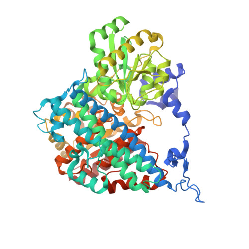
6VWY

Crystal structure of C45G/T50C D. vulgaris carbon monoxide dehydrogenase (anaerobic)


Experimental Data Snapshot

  • Method: X-RAY DIFFRACTION
  • Resolution: 1.83 Å
  • R-Value Free: 0.178 
  • R-Value Work: 0.147 
  • R-Value Observed: 0.149 

wwPDB Validation   3D Report Full Report

Currently 6VWY does not have a validation slider image.


This is version 1.1 of the entry. See complete history


Literature

The Solvent-Exposed Fe-S D-Cluster Contributes to Oxygen-Resistance inDesulfovibrio vulgarisNi-Fe Carbon Monoxide Dehydrogenase.

Wittenborn, E.C.Guendon, C.Merrouch, M.Benvenuti, M.Fourmond, V.Leger, C.Drennan, C.L.Dementin, S.

(2020) ACS Catal 10: 7328-7335

  • DOI: https://doi.org/10.1021/acscatal.0c00934
  • Primary Citation of Related Structures:  
    6VWY, 6VWZ, 6VX0, 6VX1

  • PubMed Abstract: 

    Ni-Fe CO-dehydrogenases (CODHs) catalyze the conversion between CO and CO 2 using a chain of Fe-S clusters to mediate long-range electron transfer. One of these clusters, the D-cluster, is surface-exposed and serves to transfer electrons between CODH and external redox partners. These enzymes tend to be extremely O 2 -sensitive and are always manipulated under strictly anaerobic conditions. However, the CODH from Desulfovibrio vulgaris (Dv) appears unique: exposure to micromolar concentrations of O 2 on the minutes-time scale only reversibly inhibits the enzyme, and full activity is recovered after reduction. Here, we examine whether this unusual property of Dv CODH results from the nature of its D-cluster, which is a [2Fe-2S] cluster, instead of the [4Fe-4S] cluster observed in all other characterized CODHs. To this aim, we produced and characterized a Dv CODH variant where the [2Fe-2S] D-cluster is replaced with a [4Fe-4S] D-cluster through mutagenesis of the D-cluster-binding sequence motif. We determined the crystal structure of this CODH variant to 1.83-Å resolution and confirmed the incorporation of a [4Fe-4S] D-cluster. We show that upon long-term O 2 -exposure, the [4Fe-4S] D-cluster degrades, whereas the [2Fe-2S] D-cluster remains intact. Crystal structures of the Dv CODH variant exposed to O 2 for increasing periods of time provide snapshots of [4Fe-4S] D-cluster degradation. We further show that the WT enzyme purified under aerobic conditions retains 30% activity relative to a fully anaerobic purification, compared to 10% for the variant, and the WT enzyme loses activity more slowly than the variant upon prolonged aerobic storage. The D-cluster is therefore a key site of irreversible oxidative damage in Dv CODH, and the presence of a [2Fe-2S] D-cluster contributes to the O 2 -tolerance of this enzyme. Together, these results relate O 2 -sensitivity with the details of the protein structure in this family of enzymes.


  • Organizational Affiliation

    Department of Chemistry, Department of Biology, and Howard Hughes Medical Institute, Massachusetts Institute of Technology, Cambridge, Massachusetts 02139, United States.


Macromolecules
Find similar proteins by:  (by identity cutoff)  |  3D Structure
Entity ID: 1
MoleculeChains Sequence LengthOrganismDetailsImage
Carbon monoxide dehydrogenase
A, B
629Nitratidesulfovibrio vulgaris str. HildenboroughMutation(s): 2 
Gene Names: cooSDVU_2098
EC: 1.2.7.4
UniProt
Find proteins for Q72A99 (Desulfovibrio vulgaris (strain ATCC 29579 / DSM 644 / NCIMB 8303 / VKM B-1760 / Hildenborough))
Explore Q72A99 
Go to UniProtKB:  Q72A99
Entity Groups  
Sequence Clusters30% Identity50% Identity70% Identity90% Identity95% Identity100% Identity
UniProt GroupQ72A99
Sequence Annotations
Expand
  • Reference Sequence
Small Molecules
Ligands 4 Unique
IDChains Name / Formula / InChI Key2D Diagram3D Interactions
CUV
Query on CUV

Download Ideal Coordinates CCD File 
F [auth A],
L [auth B]
Fe(4)-Ni(1)-S(4) cluster, oxidized
Fe4 Ni S4
QGLWBXDZIHZONR-UHFFFAOYSA-N
XCC
Query on XCC

Download Ideal Coordinates CCD File 
E [auth A],
K [auth B]
FE(4)-NI(1)-S(4) CLUSTER
Fe4 Ni S4
QGLWBXDZIHZONR-UHFFFAOYSA-N
SF4 (Subject of Investigation/LOI)
Query on SF4

Download Ideal Coordinates CCD File 
C [auth A],
D [auth A],
J [auth B]
IRON/SULFUR CLUSTER
Fe4 S4
LJBDFODJNLIPKO-UHFFFAOYSA-N
GOL
Query on GOL

Download Ideal Coordinates CCD File 
G [auth A],
H [auth A],
I [auth A],
M [auth B],
N [auth B]
GLYCEROL
C3 H8 O3
PEDCQBHIVMGVHV-UHFFFAOYSA-N
Experimental Data & Validation

Experimental Data

  • Method: X-RAY DIFFRACTION
  • Resolution: 1.83 Å
  • R-Value Free: 0.178 
  • R-Value Work: 0.147 
  • R-Value Observed: 0.149 
  • Space Group: P 1 21 1
Unit Cell:
Length ( Å )Angle ( ˚ )
a = 65.56α = 90
b = 147.36β = 110.78
c = 66.05γ = 90
Software Package:
Software NamePurpose
PHENIXrefinement
PDB_EXTRACTdata extraction
XDSdata reduction
XSCALEdata scaling
PHASERphasing

Structure Validation

View Full Validation Report

Currently 6VWY does not have a validation slider image.


Entry History & Funding Information

Deposition Data


Funding OrganizationLocationGrant Number
National Institutes of Health/National Institute of General Medical Sciences (NIH/NIGMS)United StatesR01 GM069857
National Institutes of Health/National Institute of General Medical Sciences (NIH/NIGMS)United StatesR35 GM126982
National Institutes of Health/National Institute of General Medical Sciences (NIH/NIGMS)United StatesT32 GM008334
Howard Hughes Medical Institute (HHMI)United States--

Revision History  (Full details and data files)

  • Version 1.0: 2020-07-29
    Type: Initial release
  • Version 1.1: 2023-10-11
    Changes: Data collection, Database references, Refinement description