6VT9

Naegleria gruberi RNA ligase E227A mutant with ATP and Mn


Experimental Data Snapshot

  • Method: X-RAY DIFFRACTION
  • Resolution: 2.49 Å
  • R-Value Free: 0.305 
  • R-Value Work: 0.219 
  • R-Value Observed: 0.227 

wwPDB Validation   3D Report Full Report

Currently 6VT9 does not have a validation slider image.


This is version 1.3 of the entry. See complete history


Literature

Caveat mutator: alanine substitutions for conserved amino acids in RNA ligase elicit unexpected rearrangements of the active site for lysine adenylylation.

Unciuleac, M.C.Goldgur, Y.Shuman, S.

(2020) Nucleic Acids Res 48: 5603-5615

  • DOI: https://doi.org/10.1093/nar/gkaa238
  • Primary Citation of Related Structures:  
    6VT0, 6VT1, 6VT3, 6VT4, 6VT5, 6VT6, 6VT8, 6VT9, 6VTB, 6VTD, 6VTE, 6VTF, 6VTG

  • PubMed Abstract: 

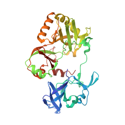
    Naegleria gruberi RNA ligase (NgrRnl) exemplifies the Rnl5 family of adenosine triphosphate (ATP)-dependent polynucleotide ligases that seal 3'-OH RNA strands in the context of 3'-OH/5'-PO4 nicked duplexes. Like all classic ligases, NgrRnl forms a covalent lysyl-AMP intermediate. A two-metal mechanism of lysine adenylylation was established via a crystal structure of the NgrRnl•ATP•(Mn2+)2 Michaelis complex. Here we conducted an alanine scan of active site constituents that engage the ATP phosphates and the metal cofactors. We then determined crystal structures of ligase-defective NgrRnl-Ala mutants in complexes with ATP/Mn2+. The unexpected findings were that mutations K170A, E227A, K326A and R149A (none of which impacted overall enzyme structure) triggered adverse secondary changes in the active site entailing dislocations of the ATP phosphates, altered contacts to ATP, and variations in the numbers and positions of the metal ions that perverted the active sites into off-pathway states incompatible with lysine adenylylation. Each alanine mutation elicited a distinctive off-pathway distortion of the ligase active site. Our results illuminate a surprising plasticity of the ligase active site in its interactions with ATP and metals. More broadly, they underscore a valuable caveat when interpreting mutational data in the course of enzyme structure-function studies.


  • Organizational Affiliation

    Molecular Biology, Sloan-Kettering Institute, 1275 York Avenue, New York, NY 10065, USA.


Macromolecules
Find similar proteins by:  (by identity cutoff)  |  3D Structure
Entity ID: 1
MoleculeChains Sequence LengthOrganismDetailsImage
RNA ligase340Naegleria gruberiMutation(s): 1 
Gene Names: NAEGRDRAFT_82186
UniProt
Find proteins for D2W2Z5 (Naegleria gruberi)
Explore D2W2Z5 
Go to UniProtKB:  D2W2Z5
Entity Groups  
Sequence Clusters30% Identity50% Identity70% Identity90% Identity95% Identity100% Identity
UniProt GroupD2W2Z5
Sequence Annotations
Expand
  • Reference Sequence
Experimental Data & Validation

Experimental Data

  • Method: X-RAY DIFFRACTION
  • Resolution: 2.49 Å
  • R-Value Free: 0.305 
  • R-Value Work: 0.219 
  • R-Value Observed: 0.227 
  • Space Group: P 41 21 2
Unit Cell:
Length ( Å )Angle ( ˚ )
a = 88.432α = 90
b = 88.432β = 90
c = 105.347γ = 90
Software Package:
Software NamePurpose
SCALEPACKdata scaling
PHENIXrefinement
PDB_EXTRACTdata extraction
HKL-2000data reduction
PHENIXphasing

Structure Validation

View Full Validation Report

Currently 6VT9 does not have a validation slider image.


Entry History & Funding Information

Deposition Data


Funding OrganizationLocationGrant Number
National Institutes of Health/National Institute of General Medical Sciences (NIH/NIGMS)United StatesR35-GM126945

Revision History  (Full details and data files)

  • Version 1.0: 2020-04-08
    Type: Initial release
  • Version 1.1: 2020-05-06
    Changes: Database references
  • Version 1.2: 2020-06-10
    Changes: Database references
  • Version 1.3: 2023-10-11
    Changes: Data collection, Database references, Refinement description