Cellular and Structural Basis of Synthesis of the Unique Intermediate Dehydro-F420-0 in Mycobacteria.
Grinter, R., Ney, B., Brammananth, R., Barlow, C.K., Cordero, P.R.F., Gillett, D.L., Izore, T., Cryle, M.J., Harold, L.K., Cook, G.M., Taiaroa, G., Williamson, D.A., Warden, A.C., Oakeshott, J.G., Taylor, M.C., Crellin, P.K., Jackson, C.J., Schittenhelm, R.B., Coppel, R.L., Greening, C.(2020) mSystems 5
- PubMed: 32430409
- DOI: https://doi.org/10.1128/mSystems.00389-20
- Primary Citation of Related Structures:
6UVX, 6UW1, 6UW3, 6UW5, 6UW7 - PubMed Abstract:
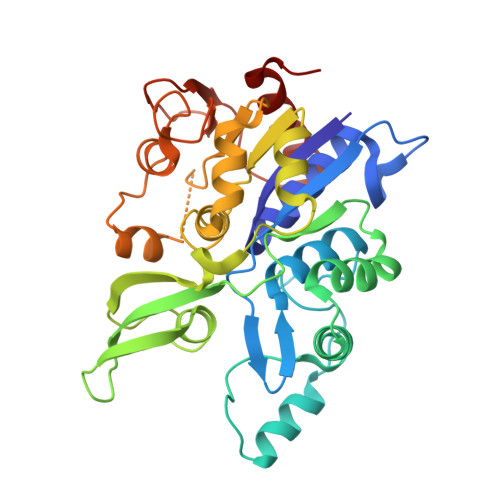
F 420 is a low-potential redox cofactor used by diverse bacteria and archaea. In mycobacteria, this cofactor has multiple roles, including adaptation to redox stress, cell wall biosynthesis, and activation of the clinical antitubercular prodrugs pretomanid and delamanid. A recent biochemical study proposed a revised biosynthesis pathway for F 420 in mycobacteria; it was suggested that phosphoenolpyruvate served as a metabolic precursor for this pathway, rather than 2-phospholactate as long proposed, but these findings were subsequently challenged. In this work, we combined metabolomic, genetic, and structural analyses to resolve these discrepancies and determine the basis of F 420 biosynthesis in mycobacterial cells. We show that, in whole cells of Mycobacterium smegmatis , phosphoenolpyruvate rather than 2-phospholactate stimulates F 420 biosynthesis. Analysis of F 420 biosynthesis intermediates present in M. smegmatis cells harboring genetic deletions at each step of the biosynthetic pathway confirmed that phosphoenolpyruvate is then used to produce the novel precursor compound dehydro-F 420 -0. To determine the structural basis of dehydro-F 420 -0 production, we solved high-resolution crystal structures of the enzyme responsible (FbiA) in apo-, substrate-, and product-bound forms. These data show the essential role of a single divalent cation in coordinating the catalytic precomplex of this enzyme and demonstrate that dehydro-F 420 -0 synthesis occurs through a direct substrate transfer mechanism. Together, these findings resolve the biosynthetic pathway of F 420 in mycobacteria and have significant implications for understanding the emergence of antitubercular prodrug resistance. IMPORTANCE Mycobacteria are major environmental microorganisms and cause many significant diseases, including tuberculosis. Mycobacteria make an unusual vitamin-like compound, F 420 , and use it to both persist during stress and resist antibiotic treatment. Understanding how mycobacteria make F 420 is important, as this process can be targeted to create new drugs to combat infections like tuberculosis. In this study, we show that mycobacteria make F 420 in a way that is different from other bacteria. We studied the molecular machinery that mycobacteria use to make F 420 , determining the chemical mechanism for this process and identifying a novel chemical intermediate. These findings also have clinical relevance, given that two new prodrugs for tuberculosis treatment are activated by F 420 .
Organizational Affiliation:
School of Biological Sciences, Monash University, Clayton, VIC, Australia rhys.grinter@monash.edu chris.greening@monash.edu.