6U8D

Crystal structure of hepatitis C virus IRES junction IIIabc in complex with Fab HCV2


Experimental Data Snapshot

  • Method: X-RAY DIFFRACTION
  • Resolution: 1.81 Å
  • R-Value Free: 0.209 
  • R-Value Work: 0.173 
  • R-Value Observed: 0.175 

wwPDB Validation   3D Report Full Report

Currently 6U8D does not have a validation slider image.


This is version 1.3 of the entry. See complete history


Literature

Synthetic Antibody Binding to a Preorganized RNA Domain of Hepatitis C Virus Internal Ribosome Entry Site Inhibits Translation.

Koirala, D.Lewicka, A.Koldobskaya, Y.Huang, H.Piccirilli, J.A.

(2020) ACS Chem Biol 15: 205-216

  • DOI: https://doi.org/10.1021/acschembio.9b00785
  • Primary Citation of Related Structures:  
    6U8D, 6U8K

  • PubMed Abstract: 

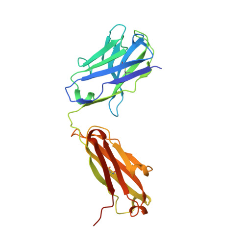
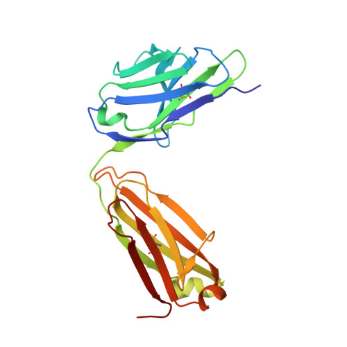
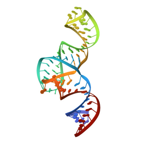
    Structured RNA elements within the internal ribosome entry site (IRES) of hepatitis C virus (HCV) genome hijack host cell machinery for translation initiation through a cap-independent mechanism. Here, using a phage display selection, we obtained two antibody fragments (Fabs), HCV2 and HCV3, against HCV IRES that bind the RNA with dissociation constants of 32 ± 7 nM and 37 ± 8 nM respectively, specifically recognizing the so-called junction IIIabc (JIIIabc). We used these Fabs as crystallization chaperones and determined the high-resolution crystal structures of JIIIabc-HCV2 and -HCV3 complexes at 1.81 Å and 2.75 Å resolution respectively, revealing an antiparallel four-way junction with the IIIa and IIIc subdomains brought together through tertiary interactions. The RNA conformation observed in the structures supports the structural model for this region derived from cryo-EM data for the HCV IRES-40S ribosome complex, suggesting that the tertiary fold of the RNA preorganizes the domain for interactions with the 40S ribosome. Strikingly, both Fabs and the ribosomal protein eS27 not only interact with a common subset of nucleotides within the JIIIabc but also use physiochemically similar sets of protein residues to do so, suggesting that the RNA surface is well-suited for interactions with proteins, perhaps analogous to the "hot spot" concept elaborated for protein-protein interactions. Using a rabbit reticulocyte lysate-based translation assay with a bicistronic reporter construct, we further demonstrated that Fabs HCV2 and HCV3 specifically inhibit the HCV IRES-directed translation, implicating disruption of the JIIIabc-ribosome interaction as a potential therapeutic strategy against HCV.


  • Organizational Affiliation

    Department of Biochemistry and Molecular Biology , The University of Chicago , Chicago , Illinois 60637 , United States.


Macromolecules

Find similar proteins by:  (by identity cutoff)  |  3D Structure
Entity ID: 2
MoleculeChains Sequence LengthOrganismDetailsImage
Heavy chain of Fab HCV2B [auth H]233Homo sapiensMutation(s): 0 
Entity Groups  
Sequence Clusters30% Identity50% Identity70% Identity90% Identity95% Identity100% Identity
Sequence Annotations
Expand
  • Reference Sequence
Find similar proteins by:  (by identity cutoff)  |  3D Structure
Entity ID: 3
MoleculeChains Sequence LengthOrganismDetailsImage
Light chain of Fab HCV2C [auth L]215Homo sapiensMutation(s): 0 
Entity Groups  
Sequence Clusters30% Identity50% Identity70% Identity90% Identity95% Identity100% Identity
Sequence Annotations
Expand
  • Reference Sequence
Find similar nucleic acids by:  (by identity cutoff)  |  3D Structure
Entity ID: 1
MoleculeChains LengthOrganismImage
JIIIabc RNA (68-MER)68Hepacivirus hominis
Sequence Annotations
Expand
  • Reference Sequence
Experimental Data & Validation

Experimental Data

  • Method: X-RAY DIFFRACTION
  • Resolution: 1.81 Å
  • R-Value Free: 0.209 
  • R-Value Work: 0.173 
  • R-Value Observed: 0.175 
  • Space Group: C 1 2 1
Unit Cell:
Length ( Å )Angle ( ˚ )
a = 49.705α = 90
b = 161.223β = 102.66
c = 96.978γ = 90
Software Package:
Software NamePurpose
PHENIXrefinement
HKL-2000data reduction
Aimlessdata scaling
PHENIXphasing

Structure Validation

View Full Validation Report

Currently 6U8D does not have a validation slider image.


Entry History & Funding Information

Deposition Data


Funding OrganizationLocationGrant Number
National Institutes of Health/National Human Genome Research Institute (NIH/NHGRI)United StatesR01AI081987
National Institutes of Health/National Human Genome Research Institute (NIH/NHGRI)United StatesR01GM102489

Revision History  (Full details and data files)

  • Version 1.0: 2019-12-04
    Type: Initial release
  • Version 1.1: 2019-12-25
    Changes: Database references
  • Version 1.2: 2020-01-29
    Changes: Database references
  • Version 1.3: 2023-10-11
    Changes: Data collection, Database references, Refinement description