Biophysical Techniques for Distinguishing Ligand Binding Modes in Cytochrome P450 Monooxygenases.
Podgorski, M.N., Harbort, J.S., Coleman, T., Stok, J.E., Yorke, J.A., Wong, L.L., Bruning, J.B., Bernhardt, P.V., De Voss, J.J., Harmer, J.R., Bell, S.G.(2020) Biochemistry 59: 1038-1050
- PubMed: 32058707
- DOI: https://doi.org/10.1021/acs.biochem.0c00027
- Primary Citation of Related Structures:
6U30, 6U3K - PubMed Abstract:
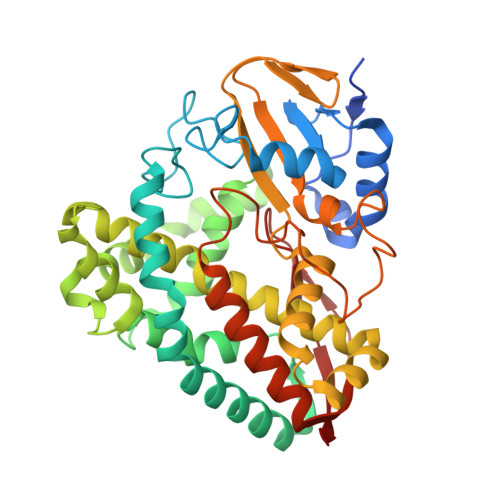
The cytochrome P450 superfamily of heme monooxygenases catalyzes important chemical reactions across nature. The changes in the optical spectra of these enzymes, induced by the addition of substrates or inhibitors, are critical for assessing how these molecules bind to the P450, enhancing or inhibiting the catalytic cycle. Here we use the bacterial CYP199A4 enzyme (Uniprot entry Q2IUO2), from Rhodopseudomonas palustris HaA2, and a range of substituted benzoic acids to investigate different binding modes. 4-Methoxybenzoic acid elicits an archetypal type I spectral response due to a ≥95% switch from the low- to high-spin state with concomitant dissociation of the sixth aqua ligand. 4-(Pyridin-3-yl)- and 4-(pyridin-2-yl)benzoic acid induced different type II ultraviolet-visible (UV-vis) spectral responses in CYP199A4. The former induced a greater red shift in the Soret wavelength (424 nm vs 422 nm) along with a larger overall absorbance change and other differences in the α-, β-, and δ-bands. There were also variations in the ferrous UV-vis spectra of these two substrate-bound forms with a spectrum indicative of Fe-N bond formation with 4-(pyridin-3-yl)benzoic acid. The crystal structures of CYP199A4, with the pyridinyl compounds bound, revealed that while the nitrogen of 4-(pyridin-3-yl)benzoic acid is coordinated to the heme, with 4-(pyridin-2-yl)benzoic acid an aqua ligand remains. Continuous wave and pulse electron paramagnetic resonance data in frozen solution revealed that the substrates are bound in the active site in a form consistent with the crystal structures. The redox potential of each CYP199A4-substrate combination was measured, allowing correlation among binding modes, spectroscopic properties, and the observed biochemical activity.
Organizational Affiliation:
Department of Chemistry, University of Adelaide, Adelaide, SA 5005, Australia.