


6TWH
Crystal structure of the haemagglutinin mutant (Gln226Leu, Gly228Ser) from an H10N7 seal influenza virus isolated in Germany
- PDB DOI: https://doi.org/10.2210/pdb6TWH/pdb
- Classification: VIRAL PROTEIN
- Organism(s): Influenza A virus (A/harbour seal/Germany/1/2014(H10N7))
- Expression System: unidentified baculovirus
- Mutation(s): Yes
- Deposited: 2020-01-13 Released: 2020-10-21
- Funding Organization(s): The Francis Crick Institute
Experimental Data Snapshot
- Method: X-RAY DIFFRACTION
- Resolution: 2.68 Å
- R-Value Free: 0.302
- R-Value Work: 0.269
- R-Value Observed: 0.271

This is version 1.1 of the entry. See complete history.

Macromolecules
Find similar proteins by:
(by identity cutoff) | 3D Structure
Entity ID: 1 | |||||
|---|---|---|---|---|---|
| Molecule | Chains | Sequence Length | Organism | Details | Image |
| Hemagglutinin | 325 | Influenza A virus (A/harbour seal/Germany/1/2014(H10N7)) | Mutation(s): 1 Gene Names: HA | 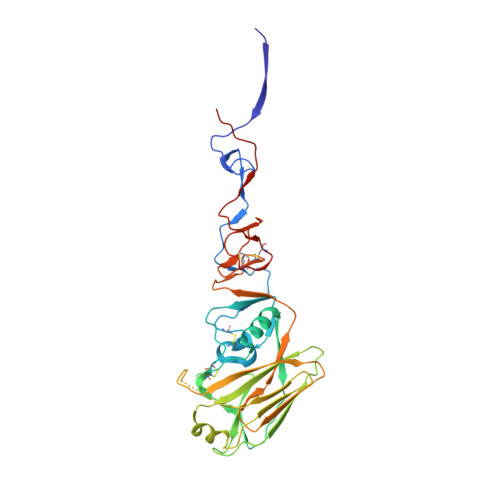 | |
UniProt | |||||
Find proteins for A0A0A7HR51 (Influenza A virus) Explore A0A0A7HR51 Go to UniProtKB: A0A0A7HR51 | |||||
Entity Groups | |||||
| Sequence Clusters | 30% Identity50% Identity70% Identity90% Identity95% Identity100% Identity | ||||
| UniProt Group | A0A0A7HR51 | ||||
Sequence AnnotationsExpand | |||||
| |||||
Find similar proteins by:
(by identity cutoff) | 3D Structure
Entity ID: 2 | |||||
|---|---|---|---|---|---|
| Molecule | Chains | Sequence Length | Organism | Details | Image |
| Hemagglutinin HA2 | 177 | Influenza A virus (A/harbour seal/Germany/1/2014(H10N7)) | Mutation(s): 0 Gene Names: HA | 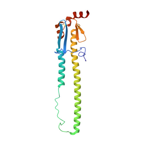 | |
UniProt | |||||
Find proteins for A0A0A7HR51 (Influenza A virus) Explore A0A0A7HR51 Go to UniProtKB: A0A0A7HR51 | |||||
Entity Groups | |||||
| Sequence Clusters | 30% Identity50% Identity70% Identity90% Identity95% Identity100% Identity | ||||
| UniProt Group | A0A0A7HR51 | ||||
Sequence AnnotationsExpand | |||||
| |||||
Oligosaccharides

Small Molecules
| Ligands 2 Unique | |||||
|---|---|---|---|---|---|
| ID | Chains | Name / Formula / InChI Key | 2D Diagram | 3D Interactions | |
| NAG Query on NAG | O [auth C] Q [auth F] R [auth F] S [auth M] T [auth N] | 2-acetamido-2-deoxy-beta-D-glucopyranose C8 H15 N O6 OVRNDRQMDRJTHS-FMDGEEDCSA-N |  | ||
| CA Query on CA | P [auth E], U [auth N], X [auth P] | CALCIUM ION Ca BHPQYMZQTOCNFJ-UHFFFAOYSA-N |  | ||
Experimental Data & Validation
Experimental Data
- Method: X-RAY DIFFRACTION
- Resolution: 2.68 Å
- R-Value Free: 0.302
- R-Value Work: 0.269
- R-Value Observed: 0.271
- Space Group: P 1 21 1
Unit Cell:
| Length ( Å ) | Angle ( ˚ ) |
|---|---|
| a = 69.31 | α = 90 |
| b = 213.61 | β = 102.29 |
| c = 156.8 | γ = 90 |
| Software Name | Purpose |
|---|---|
| REFMAC | refinement |
| PDB_EXTRACT | data extraction |
| xia2 | data reduction |
| Aimless | data scaling |
| PHASER | phasing |
Entry History & Funding Information
Deposition Data
- Released Date: 2020-10-21 Deposition Author(s): Zhang, J., Xiong, X., Purkiss, A., Walker, P., Gamblin, S., Skehel, J.J.
| Funding Organization | Location | Grant Number |
|---|---|---|
| The Francis Crick Institute | United Kingdom | -- |
Revision History (Full details and data files)
- Version 1.0: 2020-10-21
Type: Initial release - Version 1.1: 2024-01-24
Changes: Data collection, Database references, Refinement description
