6TU9

The ROR1 Pseudokinase Domain Bound To Ponatinib


Experimental Data Snapshot

  • Method: X-RAY DIFFRACTION
  • Resolution: 1.94 Å
  • R-Value Free: 0.241 
  • R-Value Work: 0.209 
  • R-Value Observed: 0.210 

wwPDB Validation   3D Report Full Report


Ligand Structure Quality Assessment 


This is version 1.3 of the entry. See complete history


Literature

Structural Insights into Pseudokinase Domains of Receptor Tyrosine Kinases.

Sheetz, J.B.Mathea, S.Karvonen, H.Malhotra, K.Chatterjee, D.Niininen, W.Perttila, R.Preuss, F.Suresh, K.Stayrook, S.E.Tsutsui, Y.Radhakrishnan, R.Ungureanu, D.Knapp, S.Lemmon, M.A.

(2020) Mol Cell 79: 390-405.e7

  • DOI: https://doi.org/10.1016/j.molcel.2020.06.018
  • Primary Citation of Related Structures:  
    6TU9, 6TUA

  • PubMed Abstract: 

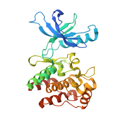
    Despite their apparent lack of catalytic activity, pseudokinases are essential signaling molecules. Here, we describe the structural and dynamic properties of pseudokinase domains from the Wnt-binding receptor tyrosine kinases (PTK7, ROR1, ROR2, and RYK), which play important roles in development. We determined structures of all pseudokinase domains in this family and found that they share a conserved inactive conformation in their activation loop that resembles the autoinhibited insulin receptor kinase (IRK). They also have inaccessible ATP-binding pockets, occluded by aromatic residues that mimic a cofactor-bound state. Structural comparisons revealed significant domain plasticity and alternative interactions that substitute for absent conserved motifs. The pseudokinases also showed dynamic properties that were strikingly similar to those of IRK. Despite the inaccessible ATP site, screening identified ATP-competitive type-II inhibitors for ROR1. Our results set the stage for an emerging therapeutic modality of "conformational disruptors" to inhibit or modulate non-catalytic functions of pseudokinases deregulated in disease.


  • Organizational Affiliation

    Department of Pharmacology, Yale University School of Medicine, New Haven, CT 06520, USA; Yale Cancer Biology Institute, Yale University West Campus, West Haven, CT 06516, USA.


Macromolecules
Find similar proteins by:  (by identity cutoff)  |  3D Structure
Entity ID: 1
MoleculeChains Sequence LengthOrganismDetailsImage
Inactive tyrosine-protein kinase transmembrane receptor ROR1
A, B
306Homo sapiensMutation(s): 0 
Gene Names: ROR1NTRKR1
UniProt & NIH Common Fund Data Resources
Find proteins for Q01973 (Homo sapiens)
Explore Q01973 
Go to UniProtKB:  Q01973
PHAROS:  Q01973
GTEx:  ENSG00000185483 
Entity Groups  
Sequence Clusters30% Identity50% Identity70% Identity90% Identity95% Identity100% Identity
UniProt GroupQ01973
Sequence Annotations
Expand
  • Reference Sequence
Small Molecules
Ligands 1 Unique
IDChains Name / Formula / InChI Key2D Diagram3D Interactions
0LI (Subject of Investigation/LOI)
Query on 0LI

Download Ideal Coordinates CCD File 
C [auth A],
D [auth B]
3-(imidazo[1,2-b]pyridazin-3-ylethynyl)-4-methyl-N-{4-[(4-methylpiperazin-1-yl)methyl]-3-(trifluoromethyl)phenyl}benzam ide
C29 H27 F3 N6 O
PHXJVRSECIGDHY-UHFFFAOYSA-N
Experimental Data & Validation

Experimental Data

  • Method: X-RAY DIFFRACTION
  • Resolution: 1.94 Å
  • R-Value Free: 0.241 
  • R-Value Work: 0.209 
  • R-Value Observed: 0.210 
  • Space Group: P 1 21 1
Unit Cell:
Length ( Å )Angle ( ˚ )
a = 54.29α = 90
b = 85.969β = 91.045
c = 72.98γ = 90
Software Package:
Software NamePurpose
PHENIXrefinement
xia2data reduction
Aimlessdata scaling
PHASERphasing

Structure Validation

View Full Validation Report


Ligand Structure Quality Assessment 


Entry History 

Deposition Data

Revision History  (Full details and data files)

  • Version 1.0: 2020-01-22
    Type: Initial release
  • Version 1.1: 2020-08-05
    Changes: Database references, Derived calculations, Structure summary
  • Version 1.2: 2020-08-19
    Changes: Database references
  • Version 1.3: 2024-01-24
    Changes: Data collection, Database references, Refinement description