Extracellular alpha/beta-hydrolase from Paenibacillus species shares structural and functional homology to tobacco salicylic acid binding protein 2.
Wilkinson, R.C., Rahman Pour, R., Jamshidi, S., Fulop, V., Bugg, T.D.H.(2020) J Struct Biol 210: 107496-107496
- PubMed: 32224091
- DOI: https://doi.org/10.1016/j.jsb.2020.107496
- Primary Citation of Related Structures:
6TJ2 - PubMed Abstract:
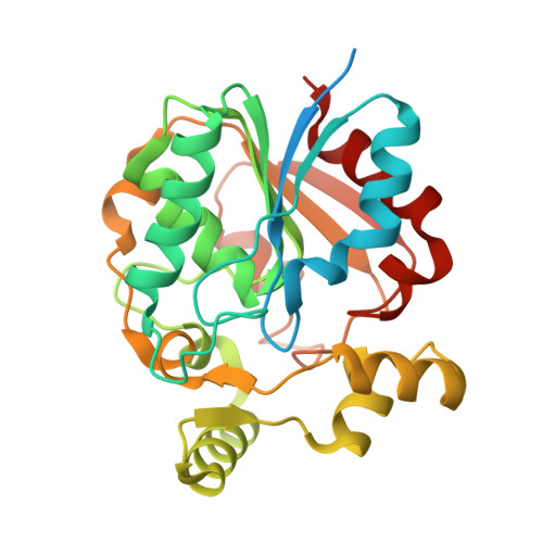
An alpha/ beta hydrolase annotated as a putative salicylate esterase within the genome of a species of Paenibacillus previously identified from differential and selective growth on Kraft lignin was structurally and functionally characterised. Feruloyl esterases are key to the degradation of lignin in several bacterial species and although this activity was investigated, no such activity was observed. The crystal structure of the Paenibacillus esterase, here denoted as PnbE, was determined at 1.32 Å resolution, showing high similarity to Nicotiana tabacum salicylic acid binding protein 2 from the protein database. Structural similarities between these two structures across the core domains and key catalytic residues were observed, with superposition of catalytic residues giving an RMSD of 0.5 Å across equivalent Cα atoms. Conversely, the cap domains of PnbE and Nicotiana tabacum SABP2 showed greater divergence with decreased flexibility in the PnbE cap structure. Activity of PnbE as a putative methyl salicylate esterase was supported with binding studies showing affinity for salicylic acid and functional studies showing methyl salicylate esterase activity. We hypothesise that this activity could enrich Paenibacillus sp. within the rhizosphere by increasing salicylic acid concentrations within the soil.
Organizational Affiliation:
School of Life Sciences, University of Warwick, Coventry CV4 7AL, UK. Electronic address: Rachael.wilkinson@path.ox.ac.uk.