6SIF

Epidermicin antimicrobial protein from Staphylococcus epidermidis


Experimental Data Snapshot

  • Method: X-RAY DIFFRACTION
  • Resolution: 1.69 Å
  • R-Value Free: 0.262 
  • R-Value Work: 0.247 
  • R-Value Observed: 0.248 

wwPDB Validation   3D Report Full Report


This is version 1.2 of the entry. See complete history


Literature

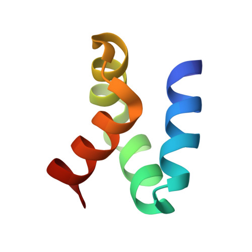
Flowering Poration-A Synergistic Multi-Mode Antibacterial Mechanism by a Bacteriocin Fold.

Hammond, K.Lewis, H.Halliwell, S.Desriac, F.Nardone, B.Ravi, J.Hoogenboom, B.W.Upton, M.Derrick, J.P.Ryadnov, M.G.

(2020) iScience 23: 101423-101423

  • DOI: https://doi.org/10.1016/j.isci.2020.101423
  • Primary Citation of Related Structures:  
    6SIF, 6SIG

  • PubMed Abstract: 

    Bacteriocins are a distinct family of antimicrobial proteins postulated to porate bacterial membranes. However, direct experimental evidence of pore formation by these proteins is lacking. Here we report a multi-mode poration mechanism induced by four-helix bacteriocins, epidermicin NI01 and aureocin A53. Using a combination of crystallography, spectroscopy, bioassays, and nanoscale imaging, we established that individual two-helix segments of epidermicin retain antibacterial activity but each of these segments adopts a particular poration mode. In the intact protein these segments act synergistically to balance out antibacterial and hemolytic activities. The study sets a precedent of multi-mode membrane disruption advancing the current understanding of structure-activity relationships in pore-forming proteins.


  • Organizational Affiliation

    National Physical Laboratory, Hampton Road, Teddington TW11 0LW, UK; London Centre for Nanotechnology, University College London, London WC1H 0AH, UK; Department of Physics & Astronomy, University College London, London WC1E 6BT, UK.


Macromolecules
Find similar proteins by:  (by identity cutoff)  |  3D Structure
Entity ID: 1
MoleculeChains Sequence LengthOrganismDetailsImage
Epidermicin locus structural protein
A, B, C, D, E
A, B, C, D, E, F, G, H
51Staphylococcus epidermidisMutation(s): 0 
UniProt
Find proteins for H9BG66 (Staphylococcus epidermidis)
Explore H9BG66 
Go to UniProtKB:  H9BG66
Entity Groups  
Sequence Clusters30% Identity50% Identity70% Identity90% Identity95% Identity100% Identity
UniProt GroupH9BG66
Sequence Annotations
Expand
  • Reference Sequence
Experimental Data & Validation

Experimental Data

  • Method: X-RAY DIFFRACTION
  • Resolution: 1.69 Å
  • R-Value Free: 0.262 
  • R-Value Work: 0.247 
  • R-Value Observed: 0.248 
  • Space Group: P 21 21 2
Unit Cell:
Length ( Å )Angle ( ˚ )
a = 92.18α = 90
b = 117.86β = 90
c = 52.93γ = 90
Software Package:
Software NamePurpose
REFMACrefinement
PDB_EXTRACTdata extraction
xia2data reduction
xia2data scaling
SHELXCDphasing

Structure Validation

View Full Validation Report


Entry History & Funding Information

Deposition Data


Funding OrganizationLocationGrant Number
Biotechnology and Biological Sciences Research CouncilUnited KingdomBB/J021474/1

Revision History  (Full details and data files)

  • Version 1.0: 2020-08-26
    Type: Initial release
  • Version 1.1: 2021-03-24
    Changes: Database references
  • Version 1.2: 2021-03-31
    Changes: Database references