6SDD

Crystal structure of D1228V cMET bound by BMS-777607


Experimental Data Snapshot

  • Method: X-RAY DIFFRACTION
  • Resolution: 1.93 Å
  • R-Value Free: 0.230 
  • R-Value Work: 0.210 
  • R-Value Observed: 0.211 

wwPDB Validation   3D Report Full Report


Ligand Structure Quality Assessment 


This is version 1.1 of the entry. See complete history


Literature

Structural and Molecular Insight into Resistance Mechanisms of First Generation cMET Inhibitors.

Collie, G.W.Koh, C.M.O'Neill, D.J.Stubbs, C.J.Khurana, P.Eddershaw, A.Snijder, A.Mauritzson, F.Barlind, L.Dale, I.L.Shaw, J.Phillips, C.Hennessy, E.J.Cheung, T.Narvaez, A.J.

(2019) ACS Med Chem Lett 10: 1322-1327

  • DOI: https://doi.org/10.1021/acsmedchemlett.9b00276
  • Primary Citation of Related Structures:  
    6SD9, 6SDC, 6SDD, 6SDE

  • PubMed Abstract: 

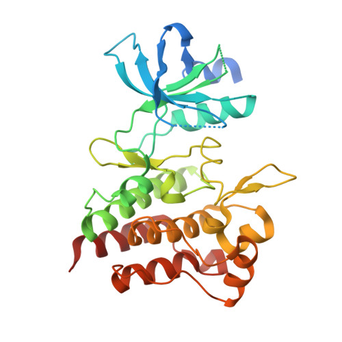
    Many small molecule inhibitors of the cMET receptor tyrosine kinase have been evaluated in clinical trials for the treatment of cancer and resistance-conferring mutations of cMET are beginning to be reported for a number of such compounds. There is now a need to understand specific cMET mutations at the molecular level, particularly concerning small molecule recognition. Toward this end, we report here the first crystal structures of the recent clinically observed resistance-conferring D1228V cMET mutant in complex with small molecule inhibitors, along with a crystal structure of wild-type cMET bound by the clinical compound savolitinib and supporting cellular, biochemical, and biophysical data. Our findings indicate that the D1228V alteration induces conformational changes in the kinase, which could have implications for small molecule inhibitor design. The data we report here increases our molecular understanding of the D1228V cMET mutation and provides insight for future inhibitor design.


  • Organizational Affiliation

    Discovery Sciences, R&D, AstraZeneca, Cambridge, U.K.


Macromolecules
Find similar proteins by:  (by identity cutoff)  |  3D Structure
Entity ID: 1
MoleculeChains Sequence LengthOrganismDetailsImage
Hepatocyte growth factor receptor309Homo sapiensMutation(s): 1 
Gene Names: MET
EC: 2.7.10.1
UniProt & NIH Common Fund Data Resources
Find proteins for P08581 (Homo sapiens)
Explore P08581 
Go to UniProtKB:  P08581
PHAROS:  P08581
GTEx:  ENSG00000105976 
Entity Groups  
Sequence Clusters30% Identity50% Identity70% Identity90% Identity95% Identity100% Identity
UniProt GroupP08581
Sequence Annotations
Expand
  • Reference Sequence
Small Molecules
Ligands 2 Unique
IDChains Name / Formula / InChI Key2D Diagram3D Interactions
353 (Subject of Investigation/LOI)
Query on 353

Download Ideal Coordinates CCD File 
B [auth A]N-{4-[(2-amino-3-chloropyridin-4-yl)oxy]-3-fluorophenyl}-4-ethoxy-1-(4-fluorophenyl)-2-oxo-1,2-dihydropyridine-3-carboxamide
C25 H19 Cl F2 N4 O4
VNBRGSXVFBYQNN-UHFFFAOYSA-N
GOL
Query on GOL

Download Ideal Coordinates CCD File 
C [auth A]GLYCEROL
C3 H8 O3
PEDCQBHIVMGVHV-UHFFFAOYSA-N
Binding Affinity Annotations 
IDSourceBinding Affinity
353 BindingDB:  6SDD IC50: min: 2.5, max: 214 (nM) from 9 assay(s)
Experimental Data & Validation

Experimental Data

  • Method: X-RAY DIFFRACTION
  • Resolution: 1.93 Å
  • R-Value Free: 0.230 
  • R-Value Work: 0.210 
  • R-Value Observed: 0.211 
  • Space Group: P 21 21 21
Unit Cell:
Length ( Å )Angle ( ˚ )
a = 57.956α = 90
b = 67.814β = 90
c = 90.047γ = 90
Software Package:
Software NamePurpose
Aimlessdata scaling
BUSTERrefinement
PDB_EXTRACTdata extraction
DIALSdata reduction
PHASERphasing

Structure Validation

View Full Validation Report


Ligand Structure Quality Assessment 


Entry History 

Deposition Data

Revision History  (Full details and data files)

  • Version 1.0: 2019-08-14
    Type: Initial release
  • Version 1.1: 2019-10-02
    Changes: Data collection, Database references