


6S08
Crystal Structure of Properdin (TSR domains N1 & 456)
- PDB DOI: https://doi.org/10.2210/pdb6S08/pdb
- Classification: IMMUNE SYSTEM
- Organism(s): Homo sapiens
- Expression System: Homo sapiens
- Mutation(s): No
- Deposited: 2019-06-14 Released: 2019-09-04
- Funding Organization(s): Dutch Kidney Foundation
Experimental Data Snapshot
- Method: X-RAY DIFFRACTION
- Resolution: 2.03 Å
- R-Value Free: 0.248
- R-Value Work: 0.212
- R-Value Observed: 0.214

This is version 2.0 of the entry. See complete history.

Macromolecules
Find similar proteins by:
(by identity cutoff) | 3D Structure
Entity ID: 1 | |||||
|---|---|---|---|---|---|
| Molecule | Chains | Sequence Length | Organism | Details | Image |
| Properdin | 225 | Homo sapiens | Mutation(s): 0 Gene Names: CFP, PFC | 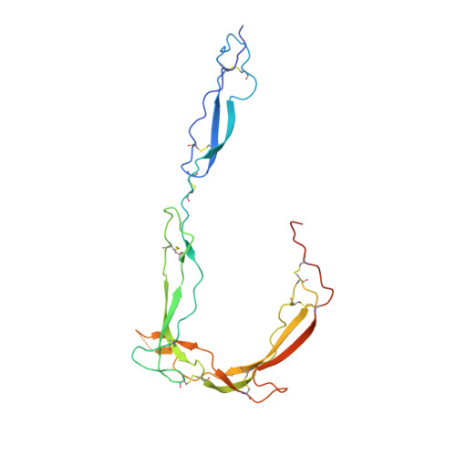 | |
UniProt & NIH Common Fund Data Resources | |||||
Find proteins for P27918 (Homo sapiens) Explore P27918 Go to UniProtKB: P27918 | |||||
PHAROS: P27918 GTEx: ENSG00000126759 | |||||
Entity Groups | |||||
| Sequence Clusters | 30% Identity50% Identity70% Identity90% Identity95% Identity100% Identity | ||||
| UniProt Group | P27918 | ||||
Sequence AnnotationsExpand | |||||
| |||||
Find similar proteins by:
(by identity cutoff) | 3D Structure
Entity ID: 2 | |||||
|---|---|---|---|---|---|
| Molecule | Chains | Sequence Length | Organism | Details | Image |
| Properdin | 110 | Homo sapiens | Mutation(s): 0 Gene Names: CFP, PFC | 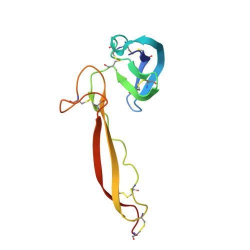 | |
UniProt & NIH Common Fund Data Resources | |||||
Find proteins for P27918 (Homo sapiens) Explore P27918 Go to UniProtKB: P27918 | |||||
PHAROS: P27918 GTEx: ENSG00000126759 | |||||
Entity Groups | |||||
| Sequence Clusters | 30% Identity50% Identity70% Identity90% Identity95% Identity100% Identity | ||||
| UniProt Group | P27918 | ||||
Sequence AnnotationsExpand | |||||
| |||||
Oligosaccharides

Small Molecules
| Ligands 4 Unique | |||||
|---|---|---|---|---|---|
| ID | Chains | Name / Formula / InChI Key | 2D Diagram | 3D Interactions | |
| MAN Query on MAN | D [auth A] E [auth A] F [auth A] G [auth A] H [auth A] | alpha-D-mannopyranose C6 H12 O6 WQZGKKKJIJFFOK-PQMKYFCFSA-N |  | ||
| FUC Query on FUC | N [auth B] | alpha-L-fucopyranose C6 H12 O5 SHZGCJCMOBCMKK-SXUWKVJYSA-N |  | ||
| PGE Query on PGE | J [auth A] | TRIETHYLENE GLYCOL C6 H14 O4 ZIBGPFATKBEMQZ-UHFFFAOYSA-N |  | ||
| NA Query on NA | L [auth A] | SODIUM ION Na FKNQFGJONOIPTF-UHFFFAOYSA-N |  | ||
Experimental Data & Validation
Experimental Data
- Method: X-RAY DIFFRACTION
- Resolution: 2.03 Å
- R-Value Free: 0.248
- R-Value Work: 0.212
- R-Value Observed: 0.214
- Space Group: C 1 2 1
Unit Cell:
| Length ( Å ) | Angle ( ˚ ) |
|---|---|
| a = 111.995 | α = 90 |
| b = 114.86 | β = 99.56 |
| c = 39.822 | γ = 90 |
| Software Name | Purpose |
|---|---|
| DIALS | data reduction |
| STARANISO | data scaling |
| PHASER | phasing |
| Coot | model building |
| REFMAC | refinement |
| PDB_EXTRACT | data extraction |
Entry History & Funding Information
Deposition Data
- Released Date: 2019-09-04 Deposition Author(s): van den Bos, R.M., Pearce, N.M., Gros, P.
| Funding Organization | Location | Grant Number |
|---|---|---|
| Dutch Kidney Foundation | Netherlands | 13OCA27 |
Revision History (Full details and data files)
- Version 1.0: 2019-09-04
Type: Initial release - Version 1.1: 2019-10-02
Changes: Data collection, Database references - Version 2.0: 2020-07-29
Type: Remediation
Reason: Carbohydrate remediation
Changes: Advisory, Atomic model, Data collection, Derived calculations, Structure summary
