6RX7

Structure of the KIV type 2 (KIV-2) domain of lipoprotein (a)


Experimental Data Snapshot

  • Method: X-RAY DIFFRACTION
  • Resolution: 1.63 Å
  • R-Value Free: 0.269 
  • R-Value Work: 0.232 
  • R-Value Observed: 0.234 

wwPDB Validation   3D Report Full Report


This is version 1.3 of the entry. See complete history


Literature

High resolution structure of human apolipoprotein (a) kringle IV type 2: beyond the lysine binding site.

Santonastaso, A.Maggi, M.De Jonge, H.Scotti, C.

(2020) J Lipid Res 61: 1687-1696

  • DOI: https://doi.org/10.1194/jlr.RA120001023
  • Primary Citation of Related Structures:  
    6RX7

  • PubMed Abstract: 

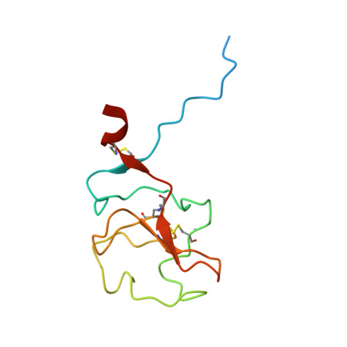
    Lipoprotein (a) [Lp(a)] is characterized by an LDL-like composition in terms of lipids and apoB100, and by one copy of a unique glycoprotein, apo(a). The apo(a) structure is mainly based on the repetition of tandem kringle domains with high homology to plasminogen kringles 4 and 5. Among them, kringle IV type 2 (KIV-2) is present in a highly variable number of genetically encoded repeats, whose length is inversely related to Lp(a) plasma concentration and cardiovascular risk. Despite it being the major component of apo(a), the actual function of KIV-2 is still unclear. Here, we describe the first high-resolution crystallographic structure of this domain. It shows a general fold very similar to other KIV domains with high and intermediate affinity for the lysine analog, ε-aminocaproic acid. Interestingly, KIV-2 presents a lysine binding site (LBS) with a unique shape and charge distribution. KIV-2 affinity for predicted small molecule binders was found to be negligible in surface plasmon resonance experiments; and with the LBS being nonfunctional, we propose to rename it "pseudo-LBS". Further investigation of the protein by computational small-molecule docking allowed us to identify a possible heparin-binding site away from the LBS, which was confirmed by specific reverse charge mutations abolishing heparin binding. This study opens new possibilities to define the pathogenesis of Lp(a)-related diseases and to facilitate the design of specific therapeutic drugs.


  • Organizational Affiliation

    Department of Molecular Medicine, Unit of Immunology and General Pathology, University of Pavia, Pavia, Italy.


Macromolecules
Find similar proteins by:  (by identity cutoff)  |  3D Structure
Entity ID: 1
MoleculeChains Sequence LengthOrganismDetailsImage
Apolipoprotein(a)
A, B, C
111Homo sapiensMutation(s): 0 
Gene Names: LPA
EC: 3.4.21
UniProt & NIH Common Fund Data Resources
Find proteins for P08519 (Homo sapiens)
Explore P08519 
Go to UniProtKB:  P08519
PHAROS:  P08519
GTEx:  ENSG00000198670 
Entity Groups  
Sequence Clusters30% Identity50% Identity70% Identity90% Identity95% Identity100% Identity
UniProt GroupP08519
Sequence Annotations
Expand
  • Reference Sequence
Small Molecules
Ligands 2 Unique
IDChains Name / Formula / InChI Key2D Diagram3D Interactions
SO4
Query on SO4

Download Ideal Coordinates CCD File 
H [auth B],
I [auth B]
SULFATE ION
O4 S
QAOWNCQODCNURD-UHFFFAOYSA-L
GOL
Query on GOL

Download Ideal Coordinates CCD File 
D [auth A]
E [auth A]
F [auth A]
G [auth A]
J [auth B]
D [auth A],
E [auth A],
F [auth A],
G [auth A],
J [auth B],
K [auth B],
L [auth B],
M [auth C]
GLYCEROL
C3 H8 O3
PEDCQBHIVMGVHV-UHFFFAOYSA-N
Experimental Data & Validation

Experimental Data

  • Method: X-RAY DIFFRACTION
  • Resolution: 1.63 Å
  • R-Value Free: 0.269 
  • R-Value Work: 0.232 
  • R-Value Observed: 0.234 
  • Space Group: C 1 2 1
Unit Cell:
Length ( Å )Angle ( ˚ )
a = 120.246α = 90
b = 42.839β = 110.23
c = 61.175γ = 90
Software Package:
Software NamePurpose
PHENIXrefinement
XDSdata reduction
Aimlessdata scaling
PHASERphasing

Structure Validation

View Full Validation Report


Entry History & Funding Information

Deposition Data


Funding OrganizationLocationGrant Number
Fondazione CARIPLOItaly42657763 (2013-0380)

Revision History  (Full details and data files)

  • Version 1.0: 2020-07-08
    Type: Initial release
  • Version 1.1: 2020-11-11
    Changes: Derived calculations
  • Version 1.2: 2021-01-20
    Changes: Database references
  • Version 1.3: 2024-01-24
    Changes: Advisory, Data collection, Database references, Refinement description