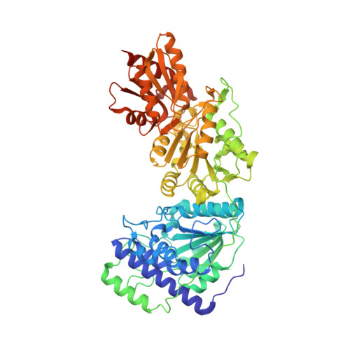
6RJB

Human transketolase variant T382E


Experimental Data Snapshot

  • Method: X-RAY DIFFRACTION
  • Resolution: 1.15 Å
  • R-Value Free: 0.148 
  • R-Value Work: 0.123 
  • R-Value Observed: 0.124 

wwPDB Validation   3D Report Full Report


Ligand Structure Quality Assessment 


This is version 2.0 of the entry. See complete history


Literature

Low-barrier hydrogen bonds in enzyme cooperativity.

Dai, S.Funk, L.M.von Pappenheim, F.R.Sautner, V.Paulikat, M.Schroder, B.Uranga, J.Mata, R.A.Tittmann, K.

(2019) Nature 573: 609-613

  • DOI: https://doi.org/10.1038/s41586-019-1581-9
  • Primary Citation of Related Structures:  
    6HA3, 6HAD, 6HAF, 6RJB, 6RJC

  • PubMed Abstract: 

    The underlying molecular mechanisms of cooperativity and allosteric regulation are well understood for many proteins, with haemoglobin and aspartate transcarbamoylase serving as prototypical examples 1,2 . The binding of effectors typically causes a structural transition of the protein that is propagated through signalling pathways to remote sites and involves marked changes on the tertiary and sometimes even the quaternary level 1-5 . However, the origin of these signals and the molecular mechanism of long-range signalling at an atomic level remain unclear 5-8 . The different spatial scales and timescales in signalling pathways render experimental observation challenging; in particular, the positions and movement of mobile protons cannot be visualized by current methods of structural analysis. Here we report the experimental observation of fluctuating low-barrier hydrogen bonds as switching elements in cooperativity pathways of multimeric enzymes. We have observed these low-barrier hydrogen bonds in ultra-high-resolution X-ray crystallographic structures of two multimeric enzymes, and have validated their assignment using computational calculations. Catalytic events at the active sites switch between low-barrier hydrogen bonds and ordinary hydrogen bonds in a circuit that consists of acidic side chains and water molecules, transmitting a signal through the collective repositioning of protons by behaving as an atomistic Newton's cradle. The resulting communication synchronizes catalysis in the oligomer. Our studies provide several lines of evidence and a working model for not only the existence of low-barrier hydrogen bonds in proteins, but also a connection to enzyme cooperativity. This finding suggests new principles of drug and enzyme design, in which sequences of residues can be purposefully included to enable long-range communication and thus the regulation of engineered biomolecules.


  • Organizational Affiliation

    Department of Molecular Enzymology, Göttingen Centre for Molecular Biosciences and Albrecht-von-Haller Institute, Georg-August University Göttingen, Göttingen, Germany.


Macromolecules
Find similar proteins by:  (by identity cutoff)  |  3D Structure
Entity ID: 1
MoleculeChains Sequence LengthOrganismDetailsImage
Transketolase
A, B
637Homo sapiensMutation(s): 1 
Gene Names: TKT
EC: 2.2.1.1
UniProt & NIH Common Fund Data Resources
Find proteins for P29401 (Homo sapiens)
Explore P29401 
Go to UniProtKB:  P29401
PHAROS:  P29401
GTEx:  ENSG00000163931 
Entity Groups  
Sequence Clusters30% Identity50% Identity70% Identity90% Identity95% Identity100% Identity
UniProt GroupP29401
Sequence Annotations
Expand
  • Reference Sequence
Small Molecules
Ligands 7 Unique
IDChains Name / Formula / InChI Key2D Diagram3D Interactions
TPP (Subject of Investigation/LOI)
Query on TPP

Download Ideal Coordinates CCD File 
C [auth A],
DA [auth B]
THIAMINE DIPHOSPHATE
C12 H19 N4 O7 P2 S
AYEKOFBPNLCAJY-UHFFFAOYSA-O
PEG
Query on PEG

Download Ideal Coordinates CCD File 
BB [auth B],
CA [auth A]
DI(HYDROXYETHYL)ETHER
C4 H10 O3
MTHSVFCYNBDYFN-UHFFFAOYSA-N
GOL
Query on GOL

Download Ideal Coordinates CCD File 
F [auth A],
GA [auth B]
GLYCEROL
C3 H8 O3
PEDCQBHIVMGVHV-UHFFFAOYSA-N
EDO
Query on EDO

Download Ideal Coordinates CCD File 
AB [auth B]
G [auth A]
H [auth A]
HA [auth B]
I [auth A]
AB [auth B],
G [auth A],
H [auth A],
HA [auth B],
I [auth A],
IA [auth B],
J [auth A],
JA [auth B],
K [auth A],
KA [auth B],
L [auth A],
LA [auth B],
M [auth A],
MA [auth B],
N [auth A],
NA [auth B],
O [auth A],
OA [auth B],
P [auth A],
PA [auth B],
Q [auth A],
QA [auth B],
R [auth A],
RA [auth B],
S [auth A],
SA [auth B],
T [auth A],
TA [auth B],
U [auth A],
UA [auth B],
V [auth A],
VA [auth B],
W [auth A],
WA [auth B],
X [auth A],
XA [auth B],
Y [auth A],
YA [auth B],
Z [auth A],
ZA [auth B]
1,2-ETHANEDIOL
C2 H6 O2
LYCAIKOWRPUZTN-UHFFFAOYSA-N
CA
Query on CA

Download Ideal Coordinates CCD File 
E [auth A],
FA [auth B]
CALCIUM ION
Ca
BHPQYMZQTOCNFJ-UHFFFAOYSA-N
MG
Query on MG

Download Ideal Coordinates CCD File 
D [auth A],
EA [auth B]
MAGNESIUM ION
Mg
JLVVSXFLKOJNIY-UHFFFAOYSA-N
NA
Query on NA

Download Ideal Coordinates CCD File 
AA [auth A],
BA [auth A]
SODIUM ION
Na
FKNQFGJONOIPTF-UHFFFAOYSA-N
Experimental Data & Validation

Experimental Data

  • Method: X-RAY DIFFRACTION
  • Resolution: 1.15 Å
  • R-Value Free: 0.148 
  • R-Value Work: 0.123 
  • R-Value Observed: 0.124 
  • Space Group: P 1 21 1
Unit Cell:
Length ( Å )Angle ( ˚ )
a = 73α = 90
b = 85.78β = 94.15
c = 92.79γ = 90
Software Package:
Software NamePurpose
PHENIXrefinement
XDSdata reduction
XSCALEdata scaling
PHENIXphasing

Structure Validation

View Full Validation Report


Ligand Structure Quality Assessment 


Entry History & Funding Information

Deposition Data


Funding OrganizationLocationGrant Number
German Research FoundationGermanyFOR 1296/TP3

Revision History  (Full details and data files)

  • Version 1.0: 2019-09-11
    Type: Initial release
  • Version 1.1: 2019-09-18
    Changes: Data collection, Database references
  • Version 1.2: 2019-10-02
    Changes: Data collection, Database references
  • Version 1.3: 2019-10-09
    Changes: Data collection, Database references
  • Version 2.0: 2021-08-04
    Changes: Atomic model, Author supporting evidence, Data collection, Derived calculations, Non-polymer description, Structure summary