6RHV

Crystal structure of mouse CD11b I-domain (CD11b-I) in complex with Staphylococcus aureus octameric bi-component leukocidin LukGH (LukH K319A mutant)


Experimental Data Snapshot

  • Method: X-RAY DIFFRACTION
  • Resolution: 2.29 Å
  • R-Value Free: 0.227 
  • R-Value Work: 0.188 
  • R-Value Observed: 0.190 

wwPDB Validation   3D Report Full Report


This is version 1.3 of the entry. See complete history


Literature

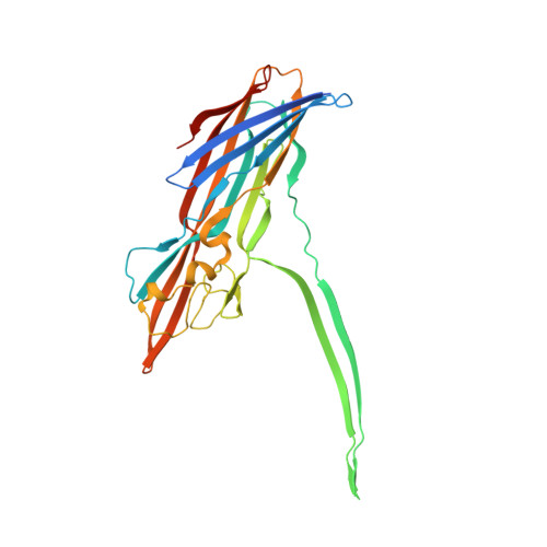
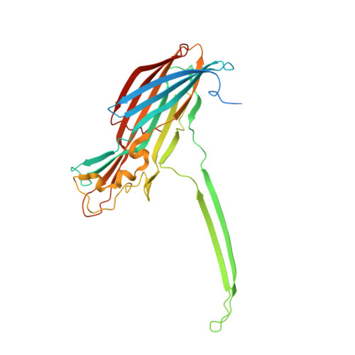
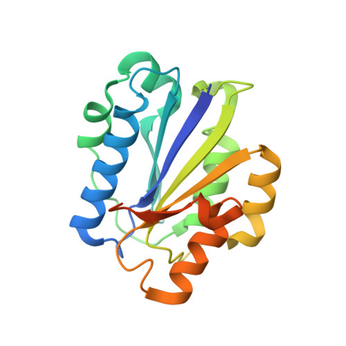
Molecular mechanism of leukocidin GH-integrin CD11b/CD18 recognition and species specificity.

Trstenjak, N.Milic, D.Graewert, M.A.Rouha, H.Svergun, D.Djinovic-Carugo, K.Nagy, E.Badarau, A.

(2020) Proc Natl Acad Sci U S A 117: 317-327

  • DOI: https://doi.org/10.1073/pnas.1913690116
  • Primary Citation of Related Structures:  
    6RHV, 6RHW

  • PubMed Abstract: 

    Host-pathogen interactions are central to understanding microbial pathogenesis. The staphylococcal pore-forming cytotoxins hijack important immune molecules but little is known about the underlying molecular mechanisms of cytotoxin-receptor interaction and host specificity. Here we report the structures of a staphylococcal pore-forming cytotoxin, leukocidin GH (LukGH), in complex with its receptor (the α-I domain of complement receptor 3, CD11b-I), both for the human and murine homologs. We observe 2 binding interfaces, on the LukG and the LukH protomers, and show that human CD11b-I induces LukGH oligomerization in solution. LukGH binds murine CD11b-I weakly and is inactive toward murine neutrophils. Using a LukGH variant engineered to bind mouse CD11b-I, we demonstrate that cytolytic activity does not only require binding but also receptor-dependent oligomerization. Our studies provide an unprecedented insight into bicomponent leukocidin-host receptor interaction, enabling the development of antitoxin approaches and improved animal models to explore these approaches.


  • Organizational Affiliation

    Arsanis Biosciences, Vienna Biocenter, 1030 Vienna, Austria.


Macromolecules
Find similar proteins by:  (by identity cutoff)  |  3D Structure
Entity ID: 1
MoleculeChains Sequence LengthOrganismDetailsImage
Beta-channel forming cytolysinA [auth G]309Staphylococcus aureusMutation(s): 0 
Gene Names: lukGELQ85_15505EP54_11070EQ90_09460HMPREF3211_02235NCTC10654_02179NCTC10702_03203NCTC13131_01350RK64_10675
Membrane Entity: Yes 
UniProt
Find proteins for Q2FWP0 (Staphylococcus aureus (strain NCTC 8325 / PS 47))
Explore Q2FWP0 
Go to UniProtKB:  Q2FWP0
Entity Groups  
Sequence Clusters30% Identity50% Identity70% Identity90% Identity95% Identity100% Identity
UniProt GroupQ2FWP0
Sequence Annotations
Expand
  • Reference Sequence
Find similar proteins by:  (by identity cutoff)  |  3D Structure
Entity ID: 2
MoleculeChains Sequence LengthOrganismDetailsImage
Beta-channel forming cytolysinB [auth H]324Staphylococcus aureusMutation(s): 1 
Gene Names: lukHBTN44_11630EP54_11065EQ90_09455HMPREF3211_02234NCTC10654_02180NCTC10702_03204NCTC13131_01351NCTC13196_01958RK64_10680
Membrane Entity: Yes 
UniProt
Find proteins for Q2FWN9 (Staphylococcus aureus (strain NCTC 8325 / PS 47))
Explore Q2FWN9 
Go to UniProtKB:  Q2FWN9
Entity Groups  
Sequence Clusters30% Identity50% Identity70% Identity90% Identity95% Identity100% Identity
UniProt GroupQ2FWN9
Sequence Annotations
Expand
  • Reference Sequence
Find similar proteins by:  (by identity cutoff)  |  3D Structure
Entity ID: 3
MoleculeChains Sequence LengthOrganismDetailsImage
Integrin alpha-M195Mus musculusMutation(s): 0 
Gene Names: Itgam
Membrane Entity: Yes 
UniProt & NIH Common Fund Data Resources
Find proteins for P05555 (Mus musculus)
Explore P05555 
Go to UniProtKB:  P05555
IMPC:  MGI:96607
Entity Groups  
Sequence Clusters30% Identity50% Identity70% Identity90% Identity95% Identity100% Identity
UniProt GroupP05555
Sequence Annotations
Expand
  • Reference Sequence
Experimental Data & Validation

Experimental Data

  • Method: X-RAY DIFFRACTION
  • Resolution: 2.29 Å
  • R-Value Free: 0.227 
  • R-Value Work: 0.188 
  • R-Value Observed: 0.190 
  • Space Group: P 4 21 2
Unit Cell:
Length ( Å )Angle ( ˚ )
a = 130.494α = 90
b = 130.494β = 90
c = 109.051γ = 90
Software Package:
Software NamePurpose
PHENIXrefinement
XDSdata reduction
XSCALEdata scaling
PHASERphasing
Cootmodel building
BUCCANEERmodel building
ARP/wARPmodel building

Structure Validation

View Full Validation Report


Entry History & Funding Information

Deposition Data


Funding OrganizationLocationGrant Number
Christian Doppler ForschungsgesellschaftAustriaCD-Laboratory for High-Content Structural Biology and Biotechnology

Revision History  (Full details and data files)

  • Version 1.0: 2019-12-18
    Type: Initial release
  • Version 1.1: 2020-01-01
    Changes: Database references
  • Version 1.2: 2020-01-15
    Changes: Database references
  • Version 1.3: 2024-01-24
    Changes: Data collection, Database references, Derived calculations, Refinement description