6RA9

Novel structural features and post-translational modifications in eukaryotic elongation factor 1A2 from Oryctolagus cuniculus


Experimental Data Snapshot

  • Method: X-RAY DIFFRACTION
  • Resolution: 2.70 Å
  • R-Value Free: 0.224 
  • R-Value Work: 0.179 
  • R-Value Observed: 0.181 

wwPDB Validation   3D Report Full Report


Ligand Structure Quality Assessment 


This is version 1.2 of the entry. See complete history


Literature

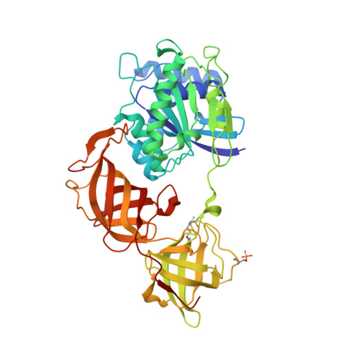
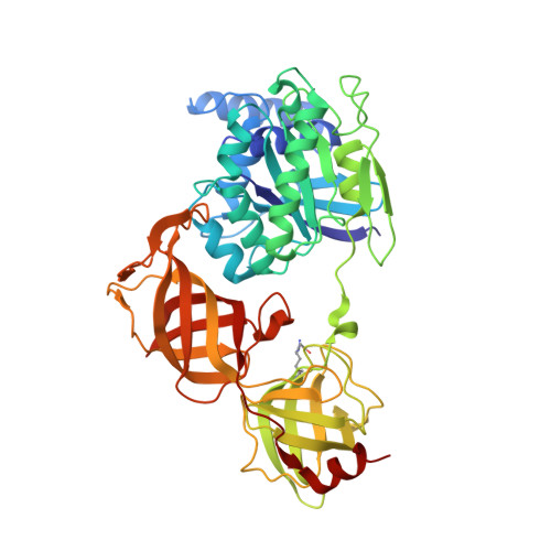
Structural Cues for Understanding eEF1A2 Moonlighting.

Carriles, A.A.Mills, A.Munoz-Alonso, M.J.Gutierrez, D.Dominguez, J.M.Hermoso, J.A.Gago, F.

(2021) Chembiochem 22: 374-391

  • DOI: https://doi.org/10.1002/cbic.202000516
  • Primary Citation of Related Structures:  
    6RA9

  • PubMed Abstract: 

    Spontaneous mutations in the EEF1A2 gene cause epilepsy and severe neurological disabilities in children. The crystal structure of eEF1A2 protein purified from rabbit skeletal muscle reveals a post-translationally modified dimer that provides information about the sites of interaction with numerous binding partners, including itself, and maps these mutations onto the dimer and tetramer interfaces. The spatial locations of the side chain carboxylates of Glu301 and Glu374, to which phosphatidylethanolamine is uniquely attached via an amide bond, define the anchoring points of eEF1A2 to cellular membranes and interorganellar membrane contact sites. Additional bioinformatic and molecular modeling results provide novel structural insight into the demonstrated binding of eEF1A2 to SH3 domains, the common MAPK docking groove, filamentous actin, and phosphatidylinositol-4 kinase IIIβ. In this new light, the role of eEF1A2 as an ancient, multifaceted, and articulated G protein at the crossroads of autophagy, oncogenesis and viral replication appears very distant from the "canonical" one of delivering aminoacyl-tRNAs to the ribosome that has dominated the scene and much of the thinking for many decades.


  • Organizational Affiliation

    Department of Crystallography and Structural Biology, Institute of Physical-Chemistry "Rocasolano" CSIC, 28006, Madrid, Spain.


Macromolecules
Find similar proteins by:  (by identity cutoff)  |  3D Structure
Entity ID: 1
MoleculeChains Sequence LengthOrganismDetailsImage
Elongation factor 1-alpha 2454Oryctolagus cuniculusMutation(s): 0 
UniProt
Find proteins for Q71V39 (Oryctolagus cuniculus)
Explore Q71V39 
Go to UniProtKB:  Q71V39
Entity Groups  
Sequence Clusters30% Identity50% Identity70% Identity90% Identity95% Identity100% Identity
UniProt GroupQ71V39
Sequence Annotations
Expand
  • Reference Sequence
Find similar proteins by:  (by identity cutoff)  |  3D Structure
Entity ID: 2
MoleculeChains Sequence LengthOrganismDetailsImage
Elongation factor 1-alpha 2461Oryctolagus cuniculusMutation(s): 0 
UniProt
Find proteins for Q71V39 (Oryctolagus cuniculus)
Explore Q71V39 
Go to UniProtKB:  Q71V39
Entity Groups  
Sequence Clusters30% Identity50% Identity70% Identity90% Identity95% Identity100% Identity
UniProt GroupQ71V39
Sequence Annotations
Expand
  • Reference Sequence
Small Molecules
Ligands 4 Unique
IDChains Name / Formula / InChI Key2D Diagram3D Interactions
GDP
Query on GDP

Download Ideal Coordinates CCD File 
E [auth A],
N [auth B]
GUANOSINE-5'-DIPHOSPHATE
C10 H15 N5 O11 P2
QGWNDRXFNXRZMB-UUOKFMHZSA-N
GPE
Query on GPE

Download Ideal Coordinates CCD File 
C [auth A],
D [auth A],
M [auth B]
L-ALPHA-GLYCEROPHOSPHORYLETHANOLAMINE
C5 H14 N O6 P
JZNWSCPGTDBMEW-RXMQYKEDSA-N
SO4
Query on SO4

Download Ideal Coordinates CCD File 
H [auth A]
I [auth A]
J [auth A]
K [auth A]
L [auth A]
H [auth A],
I [auth A],
J [auth A],
K [auth A],
L [auth A],
S [auth B],
T [auth B],
U [auth B],
V [auth B],
W [auth B],
X [auth B]
SULFATE ION
O4 S
QAOWNCQODCNURD-UHFFFAOYSA-L
GOL
Query on GOL

Download Ideal Coordinates CCD File 
F [auth A]
G [auth A]
O [auth B]
P [auth B]
Q [auth B]
F [auth A],
G [auth A],
O [auth B],
P [auth B],
Q [auth B],
R [auth B]
GLYCEROL
C3 H8 O3
PEDCQBHIVMGVHV-UHFFFAOYSA-N
Modified Residues  2 Unique
IDChains TypeFormula2D DiagramParent
POK
Query on POK
A
L-PEPTIDE LINKINGC8 H16 N4 O4ARG
SEP
Query on SEP
A
L-PEPTIDE LINKINGC3 H8 N O6 PSER
Experimental Data & Validation

Experimental Data

  • Method: X-RAY DIFFRACTION
  • Resolution: 2.70 Å
  • R-Value Free: 0.224 
  • R-Value Work: 0.179 
  • R-Value Observed: 0.181 
  • Space Group: P 61 2 2
Unit Cell:
Length ( Å )Angle ( ˚ )
a = 133.52α = 90
b = 133.52β = 90
c = 305.44γ = 120
Software Package:
Software NamePurpose
PHENIXrefinement
PHENIXrefinement
MOSFLMdata reduction
Aimlessdata scaling
MOLREPphasing

Structure Validation

View Full Validation Report


Ligand Structure Quality Assessment 


Entry History & Funding Information

Deposition Data


Funding OrganizationLocationGrant Number
Spanish Ministry of Science, Innovation, and UniversitiesSpainSAF2015-64629-C2-2-R

Revision History  (Full details and data files)

  • Version 1.0: 2020-06-03
    Type: Initial release
  • Version 1.1: 2021-04-07
    Changes: Database references
  • Version 1.2: 2024-01-24
    Changes: Data collection, Database references, Derived calculations, Refinement description