Radiation-damage investigation of a DNA 16-mer.
Bugris, V., Harmat, V., Ferenc, G., Brockhauser, S., Carmichael, I., Garman, E.F.(2019) J Synchrotron Radiat 26: 998-1009
- PubMed: 31274421
- DOI: https://doi.org/10.1107/S160057751900763X
- Primary Citation of Related Structures:
6QT1, 6QT2, 6QT3, 6QT4, 6QT5, 6QT6 - PubMed Abstract:
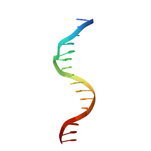
In macromolecular crystallography, a great deal of effort has been invested in understanding radiation-damage progression. While the sensitivity of protein crystals has been well characterized, crystals of DNA and of DNA-protein complexes have not thus far been studied as thoroughly. Here, a systematic investigation of radiation damage to a crystal of a DNA 16-mer diffracting to 1.8 Å resolution and held at 100 K, up to an absorbed dose of 45 MGy, is reported. The RIDL (Radiation-Induced Density Loss) automated computational tool was used for electron-density analysis. Both the global and specific damage to the DNA crystal as a function of dose were monitored, following careful calibration of the X-ray flux and beam profile. The DNA crystal was found to be fairly radiation insensitive to both global and specific damage, with half of the initial diffraction intensity being lost at an absorbed average diffraction-weighted dose, D 1/2 , of 19 MGy, compared with 9 MGy for chicken egg-white lysozyme crystals under the same beam conditions but at the higher resolution of 1.4 Å. The coefficient of sensitivity of the DNA crystal was 0.014 Å 2 MGy -1 , which is similar to that observed for proteins. These results imply that the significantly greater radiation hardness of DNA and RNA compared with protein observed in a DNA-protein complex and an RNA-protein complex could be due to scavenging action by the protein, thereby protecting the DNA and RNA in these studies. In terms of specific damage, the regions of DNA that were found to be sensitive were those associated with some of the bound calcium ions sequestered from the crystallization buffer. In contrast, moieties farther from these sites showed only small changes even at higher doses.
Organizational Affiliation:
X-ray Crystallography Laboratory, Biological Research Centre, HAC-BRC, Temesvári krt. 62, Szeged 6726, Hungary.