6QJS

Crystal structure of a DNA dodecamer containing a tetramethylpiperidinoxyl (nitroxide) spin label


Experimental Data Snapshot

  • Method: X-RAY DIFFRACTION
  • Resolution: 1.80 Å
  • R-Value Free: 0.251 
  • R-Value Work: 0.213 
  • R-Value Observed: 0.215 

wwPDB Validation   3D Report Full Report


This is version 1.2 of the entry. See complete history


Literature

2'-Alkynyl spin-labelling is a minimally perturbing tool for DNA structural analysis.

Hardwick, J.S.Haugland, M.M.El-Sagheer, A.H.Ptchelkine, D.Beierlein, F.R.Lane, A.N.Brown, T.Lovett, J.E.Anderson, E.A.

(2020) Nucleic Acids Res 48: 2830-2840

  • DOI: https://doi.org/10.1093/nar/gkaa086
  • Primary Citation of Related Structures:  
    6QJR, 6QJS

  • PubMed Abstract: 

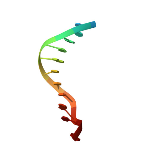
    The determination of distances between specific points in nucleic acids is essential to understanding their behaviour at the molecular level. The ability to measure distances of 2-10 nm is particularly important: deformations arising from protein binding commonly fall within this range, but the reliable measurement of such distances for a conformational ensemble remains a significant challenge. Using several techniques, we show that electron paramagnetic resonance (EPR) spectroscopy of oligonucleotides spin-labelled with triazole-appended nitroxides at the 2' position offers a robust and minimally perturbing tool for obtaining such measurements. For two nitroxides, we present results from EPR spectroscopy, X-ray crystal structures of B-form spin-labelled DNA duplexes, molecular dynamics simulations and nuclear magnetic resonance spectroscopy. These four methods are mutually supportive, and pinpoint the locations of the spin labels on the duplexes. In doing so, this work establishes 2'-alkynyl nitroxide spin-labelling as a minimally perturbing method for probing DNA conformation.


  • Organizational Affiliation

    Chemistry Research Laboratory, University of Oxford, 12 Mansfield Road, Oxford, OX1 3TA, UK.


Macromolecules

Find similar nucleic acids by:  Sequence   |   3D Structure  

Entity ID: 1
MoleculeChains LengthOrganismImage
DNA (5'-D(P*GP*CP*AP*AP*AP*TP*TP*(S6M)P*GP*CP*G)-3')
A, B
12synthetic construct
Sequence Annotations
Expand
  • Reference Sequence
Experimental Data & Validation

Experimental Data

  • Method: X-RAY DIFFRACTION
  • Resolution: 1.80 Å
  • R-Value Free: 0.251 
  • R-Value Work: 0.213 
  • R-Value Observed: 0.215 
  • Space Group: P 32 2 1
Unit Cell:
Length ( Å )Angle ( ˚ )
a = 42.999α = 90
b = 42.999β = 90
c = 97.493γ = 120
Software Package:
Software NamePurpose
REFMACrefinement
XDSdata reduction
Aimlessdata scaling
PHASERphasing

Structure Validation

View Full Validation Report


Entry History & Funding Information

Deposition Data


Funding OrganizationLocationGrant Number
Engineering and Physical Sciences Research CouncilUnited KingdomEP/M019195/1
Biotechnology and Biological Sciences Research CouncilUnited KingdomBB/J001694/1

Revision History  (Full details and data files)

  • Version 1.0: 2020-02-12
    Type: Initial release
  • Version 1.1: 2021-02-24
    Changes: Database references, Derived calculations
  • Version 1.2: 2024-01-24
    Changes: Data collection, Database references, Refinement description