6Q8V

Structure of the standard kink turn HmKt-7 variant A2bm6A.


Experimental Data Snapshot

  • Method: X-RAY DIFFRACTION
  • Resolution: 2.09 Å
  • R-Value Free: 0.211 
  • R-Value Work: 0.205 
  • R-Value Observed: 0.206 

wwPDB Validation   3D Report Full Report


This is version 1.3 of the entry. See complete history


Literature

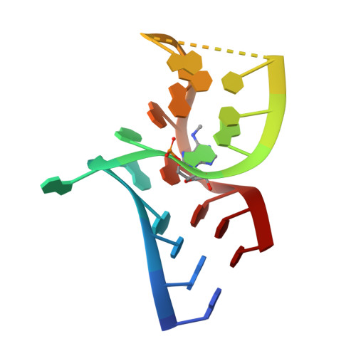
Effect of methylation of adenine N6on kink turn structure depends on location.

Ashraf, S.Huang, L.Lilley, D.M.J.

(2019) RNA Biol 16: 1377-1385

  • DOI: https://doi.org/10.1080/15476286.2019.1630797
  • Primary Citation of Related Structures:  
    6Q8U, 6Q8V

  • PubMed Abstract: 

    N 6 -methyladenine is the most common covalent modification in cellular RNA species, with demonstrated functional consequences. At the molecular level this methylation could alter local RNA structure, and/or modulate the binding of specific proteins. We have previously shown that trans -Hoogsteen-sugar (sheared) A:G base pairs can be completely disrupted by methylation, and that this occurs in a sub-set ofD/D k-turn structures. In this work we have investigated to what extent sequence context affects the severity with which inclusion of N 6 -methyladenine into different A:G base pairs of a standard k-turn affects RNA folding and L7Ae protein binding. We find that local sequence has a major influence, ranging from complete absence of folding and protein binding to a relatively mild effect. We have determined the crystal structure of one of these species both free and protein-bound, showing the environment of the methyl group and the way the modification is accommodated into the k-turn structure.


  • Organizational Affiliation

    Cancer Research UK Nucleic Acid Structure Research Group, MSI/WTB Complex, The University of Dundee , Dundee , U.K.


Macromolecules

Find similar nucleic acids by:  Sequence   |   3D Structure  

Entity ID: 1
MoleculeChains LengthOrganismImage
RNA (5'-R(*GP*GP*CP*GP*AP*AP*GP*(6MZ)P*AP*CP*CP*GP*GP*GP*GP*AP*GP*CP*C)-3')19Haloarcula marismortui
Sequence Annotations
Expand
  • Reference Sequence
Experimental Data & Validation

Experimental Data

  • Method: X-RAY DIFFRACTION
  • Resolution: 2.09 Å
  • R-Value Free: 0.211 
  • R-Value Work: 0.205 
  • R-Value Observed: 0.206 
  • Space Group: P 63 2 2
Unit Cell:
Length ( Å )Angle ( ˚ )
a = 70.66α = 90
b = 70.66β = 90
c = 48.53γ = 120
Software Package:
Software NamePurpose
PHENIXrefinement
xia2data reduction
xia2data scaling
PHASERphasing

Structure Validation

View Full Validation Report


Entry History & Funding Information

Deposition Data


Funding OrganizationLocationGrant Number
Cancer Research UKUnited KingdomA18604

Revision History  (Full details and data files)

  • Version 1.0: 2019-07-03
    Type: Initial release
  • Version 1.1: 2019-07-10
    Changes: Data collection, Database references
  • Version 1.2: 2019-09-11
    Changes: Data collection, Database references
  • Version 1.3: 2024-01-24
    Changes: Data collection, Database references, Refinement description