6PZ3

Polymerase Eta-catalyzed insertion of correct G opposite template cytarabine (AraC) residue


Experimental Data Snapshot

  • Method: X-RAY DIFFRACTION
  • Resolution: 2.40 Å
  • R-Value Free: 0.216 
  • R-Value Work: 0.174 
  • R-Value Observed: 0.176 

wwPDB Validation   3D Report Full Report


Ligand Structure Quality Assessment 


This is version 1.2 of the entry. See complete history


Literature

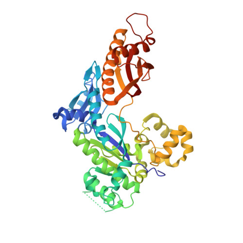
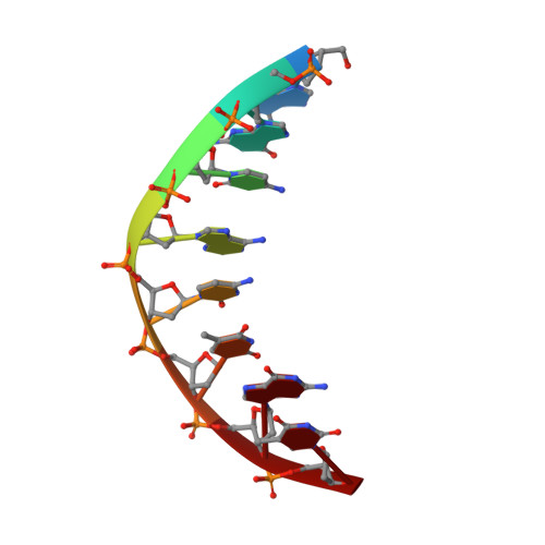
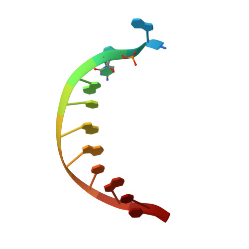
Structural insights into mutagenicity of anticancer nucleoside analog cytarabine during replication by DNA polymerase eta.

Rechkoblit, O.Johnson, R.E.Buku, A.Prakash, L.Prakash, S.Aggarwal, A.K.

(2019) Sci Rep 9: 16400-16400

  • DOI: https://doi.org/10.1038/s41598-019-52703-7
  • Primary Citation of Related Structures:  
    6PZ3, 6Q02

  • PubMed Abstract: 

    Cytarabine (AraC) is the mainstay chemotherapy for acute myeloid leukemia (AML). Whereas initial treatment with AraC is usually successful, most AML patients tend to relapse, and AraC treatment-induced mutagenesis may contribute to the development of chemo-resistant leukemic clones. We show here that whereas the high-fidelity replicative polymerase Polδ is blocked in the replication of AraC, the lower-fidelity translesion DNA synthesis (TLS) polymerase Polη is proficient, inserting both correct and incorrect nucleotides opposite a template AraC base. Furthermore, we present high-resolution crystal structures of human Polη with a template AraC residue positioned opposite correct (G) and incorrect (A) incoming deoxynucleotides. We show that Polη can accommodate local perturbation caused by the AraC via specific hydrogen bonding and maintain a reaction-ready active site alignment for insertion of both correct and incorrect incoming nucleotides. Taken together, the structures provide a novel basis for the ability of Polη to promote AraC induced mutagenesis in relapsed AML patients.


  • Organizational Affiliation

    Department of Pharmacological Sciences, Icahn School of Medicine at Mount Sinai, Box 1677, 1425 Madison Avenue, New York, NY, 10029, USA.


Macromolecules

Find similar proteins by:  (by identity cutoff)  |  3D Structure
Entity ID: 1
MoleculeChains Sequence LengthOrganismDetailsImage
DNA polymerase eta435Homo sapiensMutation(s): 1 
Gene Names: POLHRAD30RAD30AXPV
EC: 2.7.7.7
UniProt & NIH Common Fund Data Resources
Find proteins for Q9Y253 (Homo sapiens)
Explore Q9Y253 
Go to UniProtKB:  Q9Y253
PHAROS:  Q9Y253
GTEx:  ENSG00000170734 
Entity Groups  
Sequence Clusters30% Identity50% Identity70% Identity90% Identity95% Identity100% Identity
UniProt GroupQ9Y253
Sequence Annotations
Expand
  • Reference Sequence

Find similar nucleic acids by:  Sequence   |   3D Structure  

Entity ID: 2
MoleculeChains LengthOrganismImage
DNA Primer strandB [auth P]8synthetic construct
Sequence Annotations
Expand
  • Reference Sequence

Find similar nucleic acids by:  Sequence   |   3D Structure  

Entity ID: 3
MoleculeChains LengthOrganismImage
DNA template containing cytarabine (AraC) residueC [auth T]12synthetic construct
Sequence Annotations
Expand
  • Reference Sequence
Experimental Data & Validation

Experimental Data

  • Method: X-RAY DIFFRACTION
  • Resolution: 2.40 Å
  • R-Value Free: 0.216 
  • R-Value Work: 0.174 
  • R-Value Observed: 0.176 
  • Space Group: P 61
Unit Cell:
Length ( Å )Angle ( ˚ )
a = 98.221α = 90
b = 98.221β = 90
c = 81.348γ = 120
Software Package:
Software NamePurpose
HKL-2000data scaling
PHENIXrefinement
PDB_EXTRACTdata extraction
HKL-2000data reduction
PHENIXphasing

Structure Validation

View Full Validation Report


Ligand Structure Quality Assessment 


Entry History & Funding Information

Deposition Data


Funding OrganizationLocationGrant Number
National Institutes of Health/National Cancer Institute (NIH/NCI)United StatesCA200575

Revision History  (Full details and data files)

  • Version 1.0: 2019-11-20
    Type: Initial release
  • Version 1.1: 2019-12-04
    Changes: Author supporting evidence
  • Version 1.2: 2024-03-13
    Changes: Data collection, Database references, Derived calculations