6PXY

Crystal structure of ligand-binding domain of Pseudomonas fluorescens chemoreceptor CtaA in complex with L-alanine


Experimental Data Snapshot

  • Method: X-RAY DIFFRACTION
  • Resolution: 2.00 Å
  • R-Value Free: 0.249 
  • R-Value Work: 0.204 
  • R-Value Observed: 0.206 

wwPDB Validation   3D Report Full Report

Currently 6PXY does not have a validation slider image.


This is version 1.2 of the entry. See complete history


Literature

Broad Specificity of Amino Acid Chemoreceptor CtaA ofPseudomonas fluorescensIs Afforded by Plasticity of Its Amphipathic Ligand-Binding Pocket.

Ud-Din, A.I.M.S.Khan, M.F.Roujeinikova, A.

(2020) Mol Plant Microbe Interact 33: 612-623

  • DOI: https://doi.org/10.1094/MPMI-10-19-0277-R
  • Primary Citation of Related Structures:  
    6PXY, 6PY3, 6PY4, 6PY5, 6PYI, 6Q0F, 6Q0G

  • PubMed Abstract: 

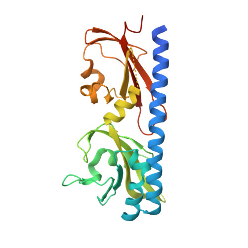
    Motile bacteria follow gradients of nutrients or other environmental cues. Many bacterial chemoreceptors that sense exogenous amino acids contain a double Cache (dCache; calcium channels and chemotaxis receptors) ligand-binding domain (LBD). A growing number of studies suggest that broad-specificity dCache-type receptors that sense more than one amino acid are common. Here, we present an investigation into the mechanism by which the dCache LBD of the chemoreceptor CtaA from a plant growth-promoting rhizobacterium, Pseudomonas fluorescens , recognizes several chemically distinct amino acids. We established that amino acids that signal by directly binding to the CtaA LBD include ones with aliphatic (l-alanine, l-proline, l-leucine, l-isoleucine, l-valine), small polar (l-serine), and large charged (l - arginine) side chains. We determined the structure of CtaA LBD in complex with different amino acids, revealing that its ability to recognize a range of structurally and chemically distinct amino acids is afforded by its easily accessible plastic pocket, which can expand or contract according to the size of the ligand side chain. The amphipathic character of the pocket enables promiscuous interactions with both polar and nonpolar amino acids. The results not only clarify the means by which various amino acids are recognized by CtaA but also reveal that a conserved mobile lid over the ligand-binding pocket adopts the same conformation in all complexes, consistent with this being an important and invariant part of the signaling mechanism.


  • Organizational Affiliation

    Infection and Immunity Program, Monash Biomedicine Discovery Institute, Australia, Department of Microbiology, Monash University, Clayton, Victoria 3800, Australia.


Macromolecules
Find similar proteins by:  (by identity cutoff)  |  3D Structure
Entity ID: 1
MoleculeChains Sequence LengthOrganismDetailsImage
Putative methyl-accepting chemotaxis protein250Pseudomonas fluorescens Pf0-1Mutation(s): 0 
Gene Names: Pfl01_4431
UniProt
Find proteins for Q3K7T6 (Pseudomonas fluorescens (strain Pf0-1))
Explore Q3K7T6 
Go to UniProtKB:  Q3K7T6
Entity Groups  
Sequence Clusters30% Identity50% Identity70% Identity90% Identity95% Identity100% Identity
UniProt GroupQ3K7T6
Sequence Annotations
Expand
  • Reference Sequence
Small Molecules
Ligands 1 Unique
IDChains Name / Formula / InChI Key2D Diagram3D Interactions
ALA (Subject of Investigation/LOI)
Query on ALA

Download Ideal Coordinates CCD File 
B [auth A]ALANINE
C3 H7 N O2
QNAYBMKLOCPYGJ-REOHCLBHSA-N
Experimental Data & Validation

Experimental Data

  • Method: X-RAY DIFFRACTION
  • Resolution: 2.00 Å
  • R-Value Free: 0.249 
  • R-Value Work: 0.204 
  • R-Value Observed: 0.206 
  • Space Group: I 21 21 21
Unit Cell:
Length ( Å )Angle ( ˚ )
a = 66.26α = 90
b = 75.28β = 90
c = 113.46γ = 90
Software Package:
Software NamePurpose
Aimlessdata scaling
REFMACrefinement
PDB_EXTRACTdata extraction
iMOSFLMdata reduction
PHASERphasing

Structure Validation

View Full Validation Report

Currently 6PXY does not have a validation slider image.


Entry History & Funding Information

Deposition Data


Funding OrganizationLocationGrant Number
Australian Research Council (ARC)Australia--

Revision History  (Full details and data files)

  • Version 1.0: 2020-03-18
    Type: Initial release
  • Version 1.1: 2020-04-15
    Changes: Database references
  • Version 1.2: 2023-10-11
    Changes: Data collection, Database references, Refinement description