6PLH

FAB fragment complexed with C-mannosylated tryptophan peptide


Experimental Data Snapshot

  • Method: X-RAY DIFFRACTION
  • Resolution: 1.60 Å
  • R-Value Free: 0.221 
  • R-Value Work: 0.189 
  • R-Value Observed: 0.191 

wwPDB Validation   3D Report Full Report


Ligand Structure Quality Assessment 


This is version 1.5 of the entry. See complete history


Literature

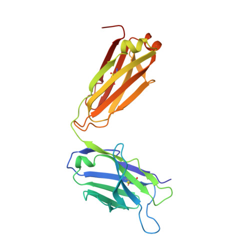
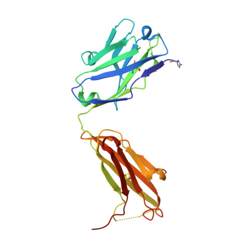
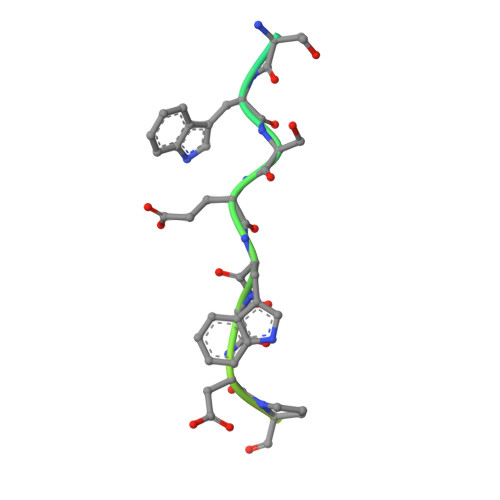
Yeast- and antibody-based tools for studying tryptophan C-mannosylation.

John, A.Jarva, M.A.Shah, S.Mao, R.Chappaz, S.Birkinshaw, R.W.Czabotar, P.E.Lo, A.W.Scott, N.E.Goddard-Borger, E.D.

(2021) Nat Chem Biol 17: 428-437

  • DOI: https://doi.org/10.1038/s41589-020-00727-w
  • Primary Citation of Related Structures:  
    6PLH

  • PubMed Abstract: 

    Tryptophan C-mannosylation is an unusual co-translational protein modification performed by metazoans and apicomplexan protists. The prevalence and biological functions of this modification are poorly understood, with progress in the field hampered by a dearth of convenient tools for installing and detecting the modification. Here, we engineer a yeast system to produce a diverse array of proteins with and without tryptophan C-mannosylation and interrogate the modification's influence on protein stability and function. This system also enabled mutagenesis studies to identify residues of the glycosyltransferase and its protein substrates that are crucial for catalysis. The collection of modified proteins accrued during this work facilitated the generation and thorough characterization of monoclonal antibodies against tryptophan C-mannosylation. These antibodies empowered proteomic analyses of the brain C-glycome by enriching for peptides possessing tryptophan C-mannosylation. This study revealed many new modification sites on proteins throughout the secretory pathway with both conventional and non-canonical consensus sequences.


  • Organizational Affiliation

    The Walter and Eliza Hall Institute of Medical Research, Parkville, Victoria, Australia.


Macromolecules
Find similar proteins by:  (by identity cutoff)  |  3D Structure
Entity ID: 1
MoleculeChains Sequence LengthOrganismDetailsImage
Fab 5G12 light chain219Mus musculusMutation(s): 0 
Entity Groups  
Sequence Clusters30% Identity50% Identity70% Identity90% Identity95% Identity100% Identity
Sequence Annotations
Expand
  • Reference Sequence
Find similar proteins by:  (by identity cutoff)  |  3D Structure
Entity ID: 2
MoleculeChains Sequence LengthOrganismDetailsImage
Fab 5G12 heavy chain223Mus musculusMutation(s): 0 
Entity Groups  
Sequence Clusters30% Identity50% Identity70% Identity90% Identity95% Identity100% Identity
Sequence Annotations
Expand
  • Reference Sequence
Find similar proteins by:  (by identity cutoff)  |  3D Structure
Entity ID: 3
MoleculeChains Sequence LengthOrganismDetailsImage
Interleukin-21 receptor37Homo sapiensMutation(s): 0 
Gene Names: IL21RNILRUNQ3121/PRO10273
UniProt & NIH Common Fund Data Resources
Find proteins for Q9HBE5 (Homo sapiens)
Explore Q9HBE5 
Go to UniProtKB:  Q9HBE5
PHAROS:  Q9HBE5
GTEx:  ENSG00000103522 
Entity Groups  
Sequence Clusters30% Identity50% Identity70% Identity90% Identity95% Identity100% Identity
UniProt GroupQ9HBE5
Sequence Annotations
Expand
  • Reference Sequence
Small Molecules
Modified Residues  1 Unique
IDChains TypeFormula2D DiagramParent
PCA
Query on PCA
B
L-PEPTIDE LINKINGC5 H7 N O3GLN
Experimental Data & Validation

Experimental Data

  • Method: X-RAY DIFFRACTION
  • Resolution: 1.60 Å
  • R-Value Free: 0.221 
  • R-Value Work: 0.189 
  • R-Value Observed: 0.191 
  • Space Group: P 1 21 1
Unit Cell:
Length ( Å )Angle ( ˚ )
a = 43.232α = 90
b = 89.892β = 95.94
c = 59.317γ = 90
Software Package:
Software NamePurpose
XDSdata reduction
Aimlessdata scaling
PHASERphasing
PHENIXrefinement
PDB_EXTRACTdata extraction

Structure Validation

View Full Validation Report


Ligand Structure Quality Assessment 


Entry History & Funding Information

Deposition Data


Funding OrganizationLocationGrant Number
National Health and Medical Research Council (NHMRC, Australia)AustraliaGNT1139546

Revision History  (Full details and data files)

  • Version 1.0: 2020-07-08
    Type: Initial release
  • Version 1.1: 2020-07-29
    Type: Remediation
    Reason: Carbohydrate remediation
    Changes: Data collection, Derived calculations, Structure summary
  • Version 1.2: 2021-02-10
    Changes: Database references, Structure summary
  • Version 1.3: 2021-02-17
    Changes: Database references
  • Version 1.4: 2021-03-31
    Changes: Database references
  • Version 1.5: 2023-10-11
    Changes: Data collection, Database references, Refinement description