6PIS

Mouse two pore domain K+ channel TRAAK (K2P4.1) - Fab complex structure


Experimental Data Snapshot

  • Method: X-RAY DIFFRACTION
  • Resolution: 2.77 Å
  • R-Value Free: 0.286 
  • R-Value Work: 0.248 
  • R-Value Observed: 0.250 

wwPDB Validation   3D Report Full Report


This is version 1.2 of the entry. See complete history


Literature

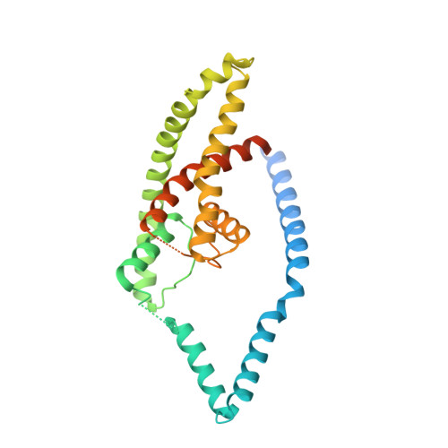
The mechanosensitive ion channel TRAAK is localized to the mammalian node of Ranvier.

Brohawn, S.G.Wang, W.Handler, A.Campbell, E.B.Schwarz, J.R.MacKinnon, R.

(2019) Elife 8

  • DOI: https://doi.org/10.7554/eLife.50403
  • Primary Citation of Related Structures:  
    6PIS

  • PubMed Abstract: 

    TRAAK is a membrane tension-activated K + channel that has been associated through behavioral studies to mechanical nociception. We used specific monoclonal antibodies in mice to show that TRAAK is localized exclusively to nodes of Ranvier, the action potential propagating elements of myelinated nerve fibers. Approximately 80 percent of myelinated nerve fibers throughout the central and peripheral nervous system contain TRAAK in what is likely an all-nodes or no-nodes per axon fashion. TRAAK is not observed at the axon initial segment where action potentials are first generated. We used polyclonal antibodies, the TRAAK inhibitor RU2 and node clamp amplifiers to demonstrate the presence and functional properties of TRAAK in rat nerve fibers. TRAAK contributes to the 'leak' K + current in mammalian nerve fiber conduction by hyperpolarizing the resting membrane potential, thereby increasing Na + channel availability for action potential propagation. We speculate on why nodes of Ranvier contain a mechanosensitive K + channel.


  • Organizational Affiliation

    Laboratory of Molecular Neurobiology and Biophysics, Howard Hughes Medical Institute, The Rockefeller University, New York, United States.


Macromolecules
Find similar proteins by:  (by identity cutoff)  |  3D Structure
Entity ID: 1
MoleculeChains Sequence LengthOrganismDetailsImage
Potassium channel subfamily K member 4
A, B
310Mus musculusMutation(s): 2 
Gene Names: Kcnk4Traak
Membrane Entity: Yes 
UniProt
Find proteins for O88454 (Mus musculus)
Explore O88454 
Go to UniProtKB:  O88454
Entity Groups  
Sequence Clusters30% Identity50% Identity70% Identity90% Identity95% Identity100% Identity
UniProt GroupO88454
Sequence Annotations
Expand
  • Reference Sequence
Find similar proteins by:  (by identity cutoff)  |  3D Structure
Entity ID: 2
MoleculeChains Sequence LengthOrganismDetailsImage
ANTIBODY FAB FRAGMENT LIGHT CHAINC [auth L],
F [auth M]
213Cricetulus migratoriusMutation(s): 0 
Entity Groups  
Sequence Clusters30% Identity50% Identity70% Identity90% Identity95% Identity100% Identity
Sequence Annotations
Expand
  • Reference Sequence
Find similar proteins by:  (by identity cutoff)  |  3D Structure
Entity ID: 3
MoleculeChains Sequence LengthOrganismDetailsImage
ANTIBODY FAB FRAGMENT HEAVY CHAIND [auth H],
E [auth I]
224Cricetulus migratoriusMutation(s): 0 
Entity Groups  
Sequence Clusters30% Identity50% Identity70% Identity90% Identity95% Identity100% Identity
Sequence Annotations
Expand
  • Reference Sequence
Experimental Data & Validation

Experimental Data

  • Method: X-RAY DIFFRACTION
  • Resolution: 2.77 Å
  • R-Value Free: 0.286 
  • R-Value Work: 0.248 
  • R-Value Observed: 0.250 
  • Space Group: P 21 21 21
Unit Cell:
Length ( Å )Angle ( ˚ )
a = 72.45α = 90
b = 154.51β = 90
c = 202.85γ = 90
Software Package:
Software NamePurpose
REFMACrefinement
XDSdata reduction
XSCALEdata scaling
PHASERphasing
PDB_EXTRACTdata extraction

Structure Validation

View Full Validation Report


Entry History & Funding Information

Deposition Data


Funding OrganizationLocationGrant Number
Howard Hughes Medical Institute (HHMI)United States--

Revision History  (Full details and data files)

  • Version 1.0: 2019-12-25
    Type: Initial release
  • Version 1.1: 2020-10-21
    Changes: Data collection, Derived calculations
  • Version 1.2: 2023-10-11
    Changes: Data collection, Database references, Refinement description