6P4F

Crystal structure of the XPB-Bax1-forked DNA ternary complex


Experimental Data Snapshot

  • Method: X-RAY DIFFRACTION
  • Resolution: 3.55 Å
  • R-Value Free: 0.272 
  • R-Value Work: 0.250 
  • R-Value Observed: 0.251 

wwPDB Validation   3D Report Full Report

Currently 6P4F does not have a validation slider image.


This is version 1.2 of the entry. See complete history


Literature

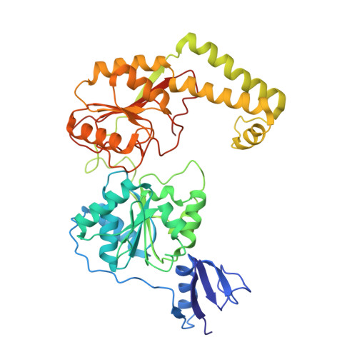
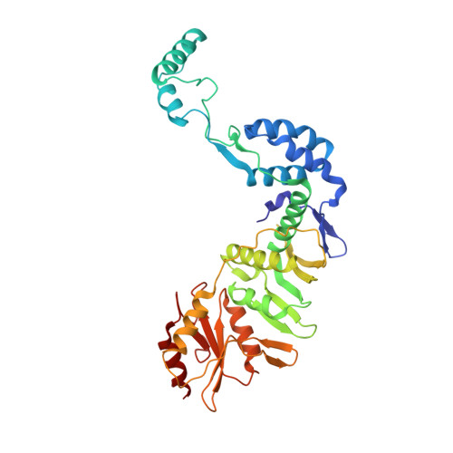
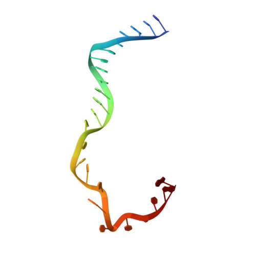
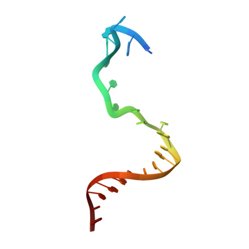
Structural basis of the XPB helicase-Bax1 nuclease complex interacting with the repair bubble DNA.

He, F.DuPrez, K.Hilario, E.Chen, Z.Fan, L.

(2020) Nucleic Acids Res 48: 11695-11705

  • DOI: https://doi.org/10.1093/nar/gkaa801
  • Primary Citation of Related Structures:  
    6P4F

  • PubMed Abstract: 

    Nucleotide excision repair (NER) removes various DNA lesions caused by UV light and chemical carcinogens. The DNA helicase XPB plays a key role in DNA opening and coordinating damage incision by nucleases during NER, but the underlying mechanisms remain unclear. Here, we report crystal structures of XPB from Sulfurisphaera tokodaii (St) bound to the nuclease Bax1 and their complex with a bubble DNA having one arm unwound in the crystal. StXPB and Bax1 together spirally encircle 10 base pairs of duplex DNA at the double-/single-stranded (ds-ss) junction. Furthermore, StXPB has its ThM motif intruding between the two DNA strands and gripping the 3'-overhang while Bax1 interacts with the 5'-overhang. This ternary complex likely reflects the state of repair bubble extension by the XPB and nuclease machine. ATP binding and hydrolysis by StXPB could lead to a spiral translocation along dsDNA and DNA strand separation by the ThM motif, revealing an unconventional DNA unwinding mechanism. Interestingly, the DNA is kept away from the nuclease domain of Bax1, potentially preventing DNA incision by Bax1 during repair bubble extension.


  • Organizational Affiliation

    Department of Biochemistry, University of California, Riverside, CA 92521, USA.


Macromolecules

Find similar proteins by:  (by identity cutoff)  |  3D Structure
Entity ID: 1
MoleculeChains Sequence LengthOrganismDetailsImage
DNA-dependent ATPase XPBII
A, C
440Sulfurisphaera tokodaii str. 7Mutation(s): 0 
Gene Names: xpb2ST1613STK_16130
UniProt
Find proteins for Q970I2 (Sulfurisphaera tokodaii (strain DSM 16993 / JCM 10545 / NBRC 100140 / 7))
Explore Q970I2 
Go to UniProtKB:  Q970I2
Entity Groups  
Sequence Clusters30% Identity50% Identity70% Identity90% Identity95% Identity100% Identity
UniProt GroupQ970I2
Sequence Annotations
Expand
  • Reference Sequence
Find similar proteins by:  (by identity cutoff)  |  3D Structure
Entity ID: 2
MoleculeChains Sequence LengthOrganismDetailsImage
Endonuclease Bax1
B, D
373Sulfurisphaera tokodaii str. 7Mutation(s): 0 
Gene Names: bax1ST1614STK_16140
UniProt
Find proteins for Q970I1 (Sulfurisphaera tokodaii (strain DSM 16993 / JCM 10545 / NBRC 100140 / 7))
Explore Q970I1 
Go to UniProtKB:  Q970I1
Entity Groups  
Sequence Clusters30% Identity50% Identity70% Identity90% Identity95% Identity100% Identity
UniProt GroupQ970I1
Sequence Annotations
Expand
  • Reference Sequence

Find similar nucleic acids by:  Sequence   |   3D Structure  

Entity ID: 3
MoleculeChains LengthOrganismImage
DNA (5'-D(*TP*TP*GP*AP*CP*TP*CP*AP*AP*CP*AP*TP*CP*CP*TP*TP*TP*GP*CP*TP*AP*CP*AP*A)-3')
E, G
24synthetic construct
Sequence Annotations
Expand
  • Reference Sequence

Find similar nucleic acids by:  Sequence   |   3D Structure  

Entity ID: 4
MoleculeChains LengthOrganismImage
DNA (5'-D(P*TP*GP*TP*AP*GP*GP*TP*TP*TP*CP*CP*AP*TP*GP*TP*TP*GP*AP*GP*TP*CP*A)-3')
F, H
23synthetic construct
Sequence Annotations
Expand
  • Reference Sequence
Small Molecules
Ligands 3 Unique
IDChains Name / Formula / InChI Key2D Diagram3D Interactions
PO4
Query on PO4

Download Ideal Coordinates CCD File 
JA [auth D],
RA [auth F],
S [auth B]
PHOSPHATE ION
O4 P
NBIIXXVUZAFLBC-UHFFFAOYSA-K
CL
Query on CL

Download Ideal Coordinates CCD File 
AA [auth B]
BA [auth B]
CA [auth B]
DA [auth B]
O [auth A]
AA [auth B],
BA [auth B],
CA [auth B],
DA [auth B],
O [auth A],
OA [auth D],
P [auth A],
Q [auth A],
R [auth A],
TA [auth G],
Z [auth B]
CHLORIDE ION
Cl
VEXZGXHMUGYJMC-UHFFFAOYSA-M
MG
Query on MG

Download Ideal Coordinates CCD File 
EA [auth C]
FA [auth C]
GA [auth C]
HA [auth C]
I [auth A]
EA [auth C],
FA [auth C],
GA [auth C],
HA [auth C],
I [auth A],
IA [auth C],
J [auth A],
K [auth A],
KA [auth D],
L [auth A],
LA [auth D],
M [auth A],
MA [auth D],
N [auth A],
NA [auth D],
PA [auth E],
QA [auth E],
SA [auth F],
T [auth B],
U [auth B],
UA [auth H],
V [auth B],
W [auth B],
X [auth B],
Y [auth B]
MAGNESIUM ION
Mg
JLVVSXFLKOJNIY-UHFFFAOYSA-N
Experimental Data & Validation

Experimental Data

  • Method: X-RAY DIFFRACTION
  • Resolution: 3.55 Å
  • R-Value Free: 0.272 
  • R-Value Work: 0.250 
  • R-Value Observed: 0.251 
  • Space Group: C 1 2 1
Unit Cell:
Length ( Å )Angle ( ˚ )
a = 214.687α = 90
b = 92.15β = 132.24
c = 172.128γ = 90
Software Package:
Software NamePurpose
MOSFLMdata reduction
SCALAdata scaling
PHASERphasing
PHENIXrefinement
PDB_EXTRACTdata extraction

Structure Validation

View Full Validation Report

Currently 6P4F does not have a validation slider image.


Entry History & Funding Information

Deposition Data


Funding OrganizationLocationGrant Number
National Institutes of Health/National Institute of General Medical Sciences (NIH/NIGMS)United StatesR01GM108893

Revision History  (Full details and data files)

  • Version 1.0: 2020-06-17
    Type: Initial release
  • Version 1.1: 2020-12-30
    Changes: Database references
  • Version 1.2: 2023-10-11
    Changes: Data collection, Database references, Refinement description