PCuAC domains from methane-oxidizing bacteria use a histidine brace to bind copper.
Fisher, O.S., Sendzik, M.R., Ross, M.O., Lawton, T.J., Hoffman, B.M., Rosenzweig, A.C.(2019) J Biol Chem 294: 16351-16363
- PubMed: 31527086
- DOI: https://doi.org/10.1074/jbc.RA119.010093
- Primary Citation of Related Structures:
6P16, 6P17, 6P1E, 6P1F, 6P1G - PubMed Abstract:
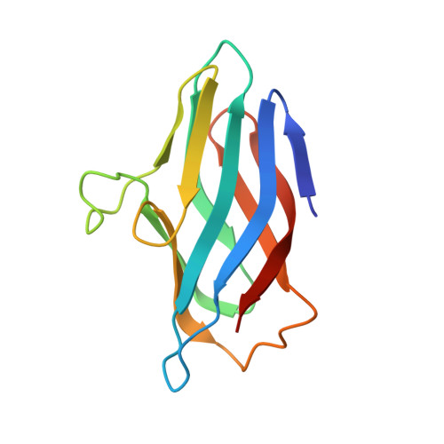
Copper is critically important for methanotrophic bacteria because their primary metabolic enzyme, particulate methane monooxygenase (pMMO), is copper-dependent. In addition to pMMO, many other copper proteins are encoded in the genomes of methanotrophs, including proteins that contain p eriplasmic c opper- A c haperone (PCu A C) domains. Using bioinformatics analyses, we identified three distinct classes of PCu A C domain-containing proteins in methanotrophs, termed PmoF1, PmoF2, and PmoF3. PCu A C domains from other types of bacteria bind a single Cu(I) ion via an H X n M X 21/22 H X M motif, which is also present in PmoF3, but PmoF1 and PmoF2 lack this motif entirely. Instead, the PCu A C domains of PmoF1 and PmoF2 bind only Cu(II), and PmoF1 binds additional Cu(II) ions in a His-rich extension to its PCu A C domain. Crystal structures of the PmoF1 and PmoF2 PCu A C domains reveal that Cu(II) is coordinated by an N-terminal histidine brace H X 10 H motif. This binding site is distinct from those of previously characterized PCu A C domains but resembles copper centers in CopC proteins and lytic polysaccharide monooxygenase (LPMO) enzymes. Bioinformatics analysis of the entire PCu A C family reveals previously unappreciated diversity, including sequences that contain both the H X n M X 21/22 H X M and H X 10 H motifs, and sequences that lack either set of copper-binding ligands. These findings provide the first characterization of an additional class of copper proteins from methanotrophs, further expand the PCu A C family, and afford new insight into the biological significance of histidine brace-mediated copper coordination.
Organizational Affiliation:
Departments of Molecular Biosciences and of Chemistry, Northwestern University, Evanston, Illinois 60208.