6OTY

Crystallographic Structure of (HbII-HbIII)-O2 from Lucina pectinata at pH 4.0


Experimental Data Snapshot

  • Method: X-RAY DIFFRACTION
  • Resolution: 2.60 Å
  • R-Value Free: 0.235 
  • R-Value Work: 0.181 
  • R-Value Observed: 0.183 

wwPDB Validation   3D Report Full Report


Ligand Structure Quality Assessment 


This is version 1.2 of the entry. See complete history


Literature

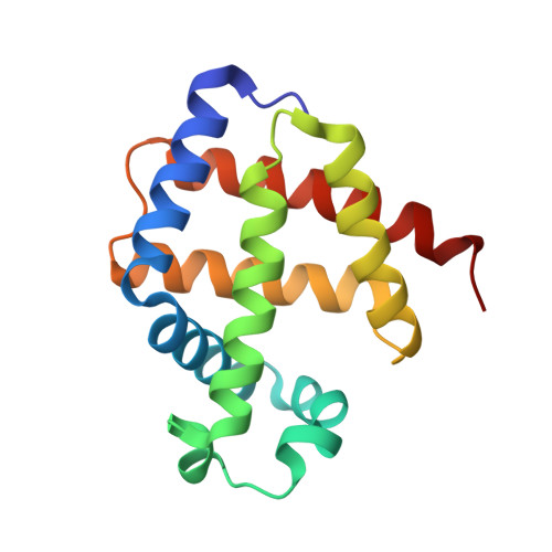
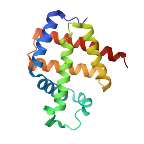
Lucina pectinata oxyhemoglobin (II-III) heterodimer pH susceptibility.

Marchany-Rivera, D.Smith, C.A.Rodriguez-Perez, J.D.Lopez-Garriga, J.

(2020) J Inorg Biochem 207: 111055-111055

  • DOI: https://doi.org/10.1016/j.jinorgbio.2020.111055
  • Primary Citation of Related Structures:  
    6OTW, 6OTX, 6OTY

  • PubMed Abstract: 

    Lucina pectinata live in high concentrations of hydrogen sulfide (H 2 S) and contains one hemoglobin, Hemoglobin I (HbI), transporting H 2 S and two hemoglobins, Hemoglobin II (HbII) and Hemoglobin (HbIII), transferring dioxygen to symbionts. HbII and HbIII contain B10 tyrosine (Tyr) and E7 glutamine (Gln) in the heme pocket generating an efficient hydrogen bonding network with the (HbII-HbIII)-O 2 species, leading to very low ligand dissociation rates. The results indicate that the oxy-hemeprotein is susceptible to pH from 4 to 9, at acidic conditions, and as a function of the potassium ferricyanide concentration, 100% of the met-aquo derivative is produced. Without a strong oxidant, pH 5 generates a small concentration of the met-aquo complex. The process is accelerated by the presence of salts, as indicated by the crystallization structures and UV-Vis spectra. The results suggest that acidic pH generates conformational changes associated with B10 and E7 heme pocket amino acids, weakening the (HbII-HbIII)-O 2 hydrogen bond network. The observation is supported by X-ray crystallography, since at pH 4 and 5, the heme-Fe tends to oxidize, while at pH 7, the oxy-heterodimer is present. Conformational changes also are observed at higher pH by the presence of a 605 nm transition associated with the iron heme-Tyr interaction. Therefore, pH is one crucial factor regulating the (HbII-HbIII)-O 2 complex hydrogen-bonding network. Thus, it can be proposed that the hydrogen bonding adjustments between the heme bound O 2 and the Tyr and Gln amino acids contribute to oxygen dissociation from the (HbII-HbIII)-O 2 system.


  • Organizational Affiliation

    Department of Chemistry, P.O. Box 9000, University of Puerto Rico, Mayagüez Campus, 00681, Puerto Rico. Electronic address: darya.marchany@upr.edu.


Macromolecules
Find similar proteins by:  (by identity cutoff)  |  3D Structure
Entity ID: 1
MoleculeChains Sequence LengthOrganismDetailsImage
Hemoglobin II152Phacoides pectinatusMutation(s): 0 
UniProt
Find proteins for P41261 (Phacoides pectinatus)
Explore P41261 
Go to UniProtKB:  P41261
Entity Groups  
Sequence Clusters30% Identity50% Identity70% Identity90% Identity95% Identity100% Identity
UniProt GroupP41261
Sequence Annotations
Expand
  • Reference Sequence
Find similar proteins by:  (by identity cutoff)  |  3D Structure
Entity ID: 2
MoleculeChains Sequence LengthOrganismDetailsImage
Hemoglobin III152Phacoides pectinatusMutation(s): 0 
UniProt
Find proteins for P41262 (Phacoides pectinatus)
Explore P41262 
Go to UniProtKB:  P41262
Entity Groups  
Sequence Clusters30% Identity50% Identity70% Identity90% Identity95% Identity100% Identity
UniProt GroupP41262
Sequence Annotations
Expand
  • Reference Sequence
Small Molecules
Experimental Data & Validation

Experimental Data

  • Method: X-RAY DIFFRACTION
  • Resolution: 2.60 Å
  • R-Value Free: 0.235 
  • R-Value Work: 0.181 
  • R-Value Observed: 0.183 
  • Space Group: P 42 21 2
Unit Cell:
Length ( Å )Angle ( ˚ )
a = 74.429α = 90
b = 74.429β = 90
c = 152.1γ = 90
Software Package:
Software NamePurpose
PHENIXrefinement
PDB_EXTRACTdata extraction
HKL-2000data reduction
HKL-2000data scaling
REFMACphasing

Structure Validation

View Full Validation Report


Ligand Structure Quality Assessment 


Entry History & Funding Information

Deposition Data


Funding OrganizationLocationGrant Number
National Science Foundation (NSF, United States)United States--

Revision History  (Full details and data files)

  • Version 1.0: 2020-04-01
    Type: Initial release
  • Version 1.1: 2020-04-08
    Changes: Database references
  • Version 1.2: 2023-10-11
    Changes: Data collection, Database references, Derived calculations, Refinement description