6OP8

S. pombe Ubc7/U7BR complex

  • Classification: LIGASE/PROTEIN BINDING
  • Organism(s): Schizosaccharomyces pombe 972h-
  • Expression System: Escherichia coli
  • Mutation(s): No 

  • Deposited: 2019-04-24 Released: 2019-08-07 
  • Deposition Author(s): Hann, Z.S., Lima, C.D.
  • Funding Organization(s): National Institutes of Health/National Institute of General Medical Sciences (NIH/NIGMS), Howard Hughes Medical Institute (HHMI), National Institutes of Health/National Cancer Institute (NIH/NCI)

Experimental Data Snapshot

  • Method: X-RAY DIFFRACTION
  • Resolution: 1.70 Å
  • R-Value Free: 0.188 
  • R-Value Work: 0.155 
  • R-Value Observed: 0.157 

wwPDB Validation   3D Report Full Report

Currently 6OP8 does not have a validation slider image.


This is version 1.3 of the entry. See complete history


Literature

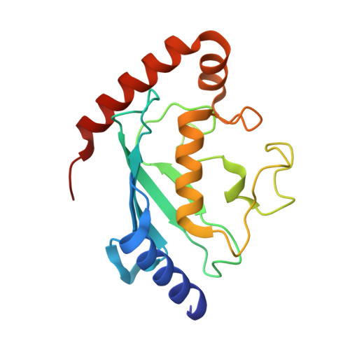
Crystal structure of the Schizosaccharomyces pombe U7BR E2-binding region in complex with Ubc7.

Hann, Z.S.Metzger, M.B.Weissman, A.M.Lima, C.D.

(2019) Acta Crystallogr F Struct Biol Commun 75: 552-560

  • DOI: https://doi.org/10.1107/S2053230X19009786
  • Primary Citation of Related Structures:  
    6OP8

  • PubMed Abstract: 

    Endoplasmic reticulum (ER)-associated degradation (ERAD) is a protein quality-control pathway in eukaryotes in which misfolded ER proteins are polyubiquitylated, extracted and ultimately degraded by the proteasome. This process involves ER membrane-embedded ubiquitin E2 and E3 enzymes, as well as a soluble E2 enzyme (Ubc7 in Saccharomyces cerevisiae and UBE2G2 in mammals). E2-binding regions (E2BRs) that recruit these soluble ERAD E2s to the ER have been identified in humans and S. cerevisiae, and structures of E2-E2BR complexes from both species have been determined. In addition to sequence and structural differences between the human and S. cerevisiae E2BRs, the binding of E2BRs also elicits different biochemical outcomes with respect to E2 charging by E1 and E2 discharge. Here, the Schizosaccharomyces pombe E2BR was identified and purified with Ubc7 to resolve a 1.7 Å resolution co-crystal structure of the E2BR in complex with Ubc7. The S. pombe E2BR binds to the back side of the E2 as an α-helix and, while differences exist, it exhibits greater similarity to the human E2BR. Structure-based sequence alignments reveal differences and conserved elements among these species. Structural comparisons and biochemistry reveal that the S. pombe E2BR presents a steric impediment to E1 binding and inhibits E1-mediated charging, respectively.


  • Organizational Affiliation

    Structural Biology Program, Sloan Kettering Institute, 1275 York Avenue, New York, NY 10065, USA.


Macromolecules
Find similar proteins by:  (by identity cutoff)  |  3D Structure
Entity ID: 1
MoleculeChains Sequence LengthOrganismDetailsImage
Ubiquitin-conjugating enzyme E2-18 kDa170Schizosaccharomyces pombe 972h-Mutation(s): 0 
Gene Names: ubc7ubcp3SPBP16F5.04
EC: 2.3.2.23
UniProt
Find proteins for O00102 (Schizosaccharomyces pombe (strain 972 / ATCC 24843))
Explore O00102 
Go to UniProtKB:  O00102
Entity Groups  
Sequence Clusters30% Identity50% Identity70% Identity90% Identity95% Identity100% Identity
UniProt GroupO00102
Sequence Annotations
Expand
  • Reference Sequence
Find similar proteins by:  (by identity cutoff)  |  3D Structure
Entity ID: 2
MoleculeChains Sequence LengthOrganismDetailsImage
CUE domain-containing protein 4, mitochondrial68Schizosaccharomyces pombe 972h-Mutation(s): 0 
Gene Names: SPCC4G3.13c
UniProt
Find proteins for P87238 (Schizosaccharomyces pombe (strain 972 / ATCC 24843))
Explore P87238 
Go to UniProtKB:  P87238
Entity Groups  
Sequence Clusters30% Identity50% Identity70% Identity90% Identity95% Identity100% Identity
UniProt GroupP87238
Sequence Annotations
Expand
  • Reference Sequence
Experimental Data & Validation

Experimental Data

  • Method: X-RAY DIFFRACTION
  • Resolution: 1.70 Å
  • R-Value Free: 0.188 
  • R-Value Work: 0.155 
  • R-Value Observed: 0.157 
  • Space Group: P 21 21 21
Unit Cell:
Length ( Å )Angle ( ˚ )
a = 26.715α = 90
b = 79.047β = 90
c = 94.521γ = 90
Software Package:
Software NamePurpose
SCALEPACKdata scaling
PHENIXrefinement
PDB_EXTRACTdata extraction
HKL-2000data reduction
PHENIXphasing

Structure Validation

View Full Validation Report

Currently 6OP8 does not have a validation slider image.


Entry History & Funding Information

Deposition Data


Funding OrganizationLocationGrant Number
National Institutes of Health/National Institute of General Medical Sciences (NIH/NIGMS)United StatesR01GM079196
National Institutes of Health/National Institute of General Medical Sciences (NIH/NIGMS)United StatesR35GM118080
Howard Hughes Medical Institute (HHMI)United States--
National Institutes of Health/National Cancer Institute (NIH/NCI)United StatesP30CA008748

Revision History  (Full details and data files)

  • Version 1.0: 2019-08-07
    Type: Initial release
  • Version 1.1: 2019-08-21
    Changes: Data collection, Database references
  • Version 1.2: 2019-11-20
    Changes: Author supporting evidence
  • Version 1.3: 2023-10-11
    Changes: Data collection, Database references, Derived calculations, Refinement description