6OOP

protein B


Experimental Data Snapshot

  • Method: X-RAY DIFFRACTION
  • Resolution: 2.80 Å

wwPDB Validation   3D Report Full Report


This is version 1.2 of the entry. See complete history


Literature

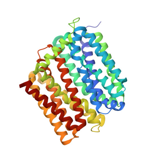
Structure of an engineered multidrug transporter MdfA reveals the molecular basis for substrate recognition.

Wu, H.H.Symersky, J.Lu, M.

(2019) Commun Biol 2: 210-210

  • DOI: https://doi.org/10.1038/s42003-019-0446-y
  • Primary Citation of Related Structures:  
    6OOM, 6OOP, 6OOQ

  • PubMed Abstract: 

    MdfA is a prototypical H + -coupled multidrug transporter that is characterized by extraordinarily broad substrate specificity. The involvement of specific H-bonds in MdfA-drug interactions and the simplicity of altering the substrate specificity of MdfA contradict the promiscuous nature of multidrug recognition, presenting a baffling conundrum. Here we show the X-ray structures of MdfA variant I239T/G354E in complexes with three electrically different ligands, determined at resolutions up to 2.2 Å. Our structures reveal that I239T/G354E interacts with these compounds differently from MdfA and that I239T/G354E possesses two discrete, non-overlapping substrate-binding sites. Our results shed new light on the molecular design of multidrug-binding and protonation sites and highlight the importance of often-neglected, long-range charge-charge interactions in multidrug recognition. Beyond helping to solve the ostensible conundrum of multidrug recognition, our findings suggest the mechanistic difference between substrate and inhibitor for any H + -dependent multidrug transporter, which may open new vistas on curtailing efflux-mediated multidrug resistance.


  • Organizational Affiliation

    Department of Biochemistry and Molecular Biology, Rosalind Franklin University of Medicine and Science, 3333 Green Bay Road, North Chicago, IL 60064 USA.


Macromolecules
Find similar proteins by:  (by identity cutoff)  |  3D Structure
Entity ID: 1
MoleculeChains Sequence LengthOrganismDetailsImage
Multidrug transporter MdfA392Escherichia coliMutation(s): 0 
Gene Names: A9R57_05385BJJ90_17655
Membrane Entity: Yes 
UniProt
Find proteins for P0AEY8 (Escherichia coli (strain K12))
Explore P0AEY8 
Go to UniProtKB:  P0AEY8
Entity Groups  
Sequence Clusters30% Identity50% Identity70% Identity90% Identity95% Identity100% Identity
UniProt GroupP0AEY8
Sequence Annotations
Expand
  • Reference Sequence
Experimental Data & Validation

Experimental Data

  • Method: X-RAY DIFFRACTION
  • Resolution: 2.80 Å
  • Space Group: C 1 2 1
Unit Cell:
Length ( Å )Angle ( ˚ )
a = 95.068α = 90
b = 63.149β = 100.87
c = 102.074γ = 90
Software Package:
Software NamePurpose
REFMACrefinement
HKL-2000data reduction
HKL-2000data scaling
SHARPphasing

Structure Validation

View Full Validation Report


Entry History & Funding Information

Deposition Data

  • Released Date: 2019-09-04 
  • Deposition Author(s): Lu, M.

Funding OrganizationLocationGrant Number
National Institutes of Health/National Institute of General Medical Sciences (NIH/NIGMS)United StatesGM094195

Revision History  (Full details and data files)

  • Version 1.0: 2019-09-04
    Type: Initial release
  • Version 1.1: 2020-01-01
    Changes: Author supporting evidence
  • Version 1.2: 2024-03-13
    Changes: Data collection, Database references, Derived calculations