6OHR

Structure of compound 5 bound human Phospholipase D1 catalytic domain


Experimental Data Snapshot

  • Method: X-RAY DIFFRACTION
  • Resolution: 3.20 Å
  • R-Value Free: 0.247 
  • R-Value Work: 0.221 
  • R-Value Observed: 0.224 

wwPDB Validation   3D Report Full Report


Ligand Structure Quality Assessment 


This is version 1.2 of the entry. See complete history


Literature

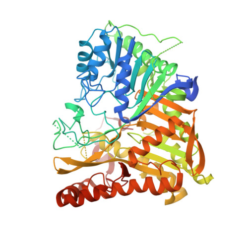
Human PLD structures enable drug design and characterization of isoenzyme selectivity.

Metrick, C.M.Peterson, E.A.Santoro, J.C.Enyedy, I.J.Murugan, P.Chen, T.Michelsen, K.Cullivan, M.Spilker, K.A.Kumar, P.R.May-Dracka, T.L.Chodaparambil, J.V.

(2020) Nat Chem Biol 16: 391-399

  • DOI: https://doi.org/10.1038/s41589-019-0458-4
  • Primary Citation of Related Structures:  
    6OHM, 6OHO, 6OHP, 6OHQ, 6OHR, 6OHS

  • PubMed Abstract: 

    Phospholipase D enzymes (PLDs) are ubiquitous phosphodiesterases that produce phosphatidic acid (PA), a key second messenger and biosynthetic building block. Although an orthologous bacterial Streptomyces sp. strain PMF PLD structure was solved two decades ago, the molecular basis underlying the functions of the human PLD enzymes (hPLD) remained unclear based on this structure due to the low homology between these sequences. Here, we describe the first crystal structures of hPLD1 and hPLD2 catalytic domains and identify novel structural elements and functional differences between the prokaryotic and eukaryotic enzymes. Furthermore, structure-based mutation studies and structures of inhibitor-hPLD complexes allowed us to elucidate the binding modes of dual and isoform-selective inhibitors, highlight key determinants of isoenzyme selectivity and provide a basis for further structure-based drug discovery and functional characterization of this therapeutically important superfamily of enzymes.


  • Organizational Affiliation

    Physical Biochemistry, Biotherapeutic and Medicinal Sciences, Biogen, Cambridge, MA, USA.


Macromolecules
Find similar proteins by:  (by identity cutoff)  |  3D Structure
Entity ID: 1
MoleculeChains Sequence LengthOrganismDetailsImage
Phospholipase D1, chimeric constuct
A, B, C, D, E
A, B, C, D, E, F
647Homo sapiensMutation(s): 0 
Gene Names: PLD1
EC: 3.1.4.4
Membrane Entity: Yes 
UniProt & NIH Common Fund Data Resources
Find proteins for Q13393 (Homo sapiens)
Explore Q13393 
Go to UniProtKB:  Q13393
PHAROS:  Q13393
GTEx:  ENSG00000075651 
Entity Groups  
Sequence Clusters30% Identity50% Identity70% Identity90% Identity95% Identity100% Identity
UniProt GroupQ13393
Sequence Annotations
Expand
  • Reference Sequence
Experimental Data & Validation

Experimental Data

  • Method: X-RAY DIFFRACTION
  • Resolution: 3.20 Å
  • R-Value Free: 0.247 
  • R-Value Work: 0.221 
  • R-Value Observed: 0.224 
  • Space Group: P 21 21 21
Unit Cell:
Length ( Å )Angle ( ˚ )
a = 93.039α = 90
b = 155.106β = 90
c = 273.56γ = 90
Software Package:
Software NamePurpose
PHENIXrefinement
XDSdata reduction
Aimlessdata scaling
PHENIXphasing

Structure Validation

View Full Validation Report


Ligand Structure Quality Assessment 


Entry History 

Deposition Data

Revision History  (Full details and data files)

  • Version 1.0: 2020-02-19
    Type: Initial release
  • Version 1.1: 2020-04-01
    Changes: Database references
  • Version 1.2: 2023-10-11
    Changes: Data collection, Database references, Refinement description