6NVM

Crystal structure of 23S rRNA methyltransferase ErmE


Experimental Data Snapshot

  • Method: X-RAY DIFFRACTION
  • Resolution: 1.75 Å
  • R-Value Free: 0.218 
  • R-Value Work: 0.192 
  • R-Value Observed: 0.194 

wwPDB Validation   3D Report Full Report


This is version 1.2 of the entry. See complete history


Literature

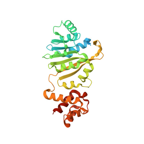
Crystal structure of ErmE - 23S rRNA methyltransferase in macrolide resistance.

Stsiapanava, A.Selmer, M.

(2019) Sci Rep 9: 14607-14607

  • DOI: https://doi.org/10.1038/s41598-019-51174-0
  • Primary Citation of Related Structures:  
    6NVM

  • PubMed Abstract: 

    Pathogens often receive antibiotic resistance genes through horizontal gene transfer from bacteria that produce natural antibiotics. ErmE is a methyltransferase (MTase) from Saccharopolyspora erythraea that dimethylates A2058 in 23S rRNA using S-adenosyl methionine (SAM) as methyl donor, protecting the ribosomes from macrolide binding. To gain insights into the mechanism of macrolide resistance, the crystal structure of ErmE was determined to 1.75 Å resolution. ErmE consists of an N-terminal Rossmann-like α/ß catalytic domain and a C-terminal helical domain. Comparison with ErmC' that despite only 24% sequence identity has the same function, reveals highly similar catalytic domains. Accordingly, superposition with the catalytic domain of ErmC' in complex with SAM suggests that the cofactor binding site is conserved. The two structures mainly differ in the C-terminal domain, which in ErmE contains a longer loop harboring an additional 3 10 helix that interacts with the catalytic domain to stabilize the tertiary structure. Notably, ErmE also differs from ErmC' by having long disordered extensions at its N- and C-termini. A C-terminal disordered region rich in arginine and glycine is also a present in two other MTases, PikR1 and PikR2, which share about 30% sequence identity with ErmE and methylate the same nucleotide in 23S rRNA.


  • Organizational Affiliation

    Department of Cell and Molecular Biology, Uppsala University, BMC, Box 596, SE-751 24, Uppsala, Sweden. alena.stsiapanava@gmail.com.


Macromolecules
Find similar proteins by:  (by identity cutoff)  |  3D Structure
Entity ID: 1
MoleculeChains Sequence LengthOrganismDetailsImage
rRNA adenine N-6-methyltransferase301Saccharopolyspora erythraeaMutation(s): 0 
Gene Names: ermESACE_0733
EC: 2.1.1.184
UniProt
Find proteins for P07287 (Saccharopolyspora erythraea (strain ATCC 11635 / DSM 40517 / JCM 4748 / NBRC 13426 / NCIMB 8594 / NRRL 2338))
Explore P07287 
Go to UniProtKB:  P07287
Entity Groups  
Sequence Clusters30% Identity50% Identity70% Identity90% Identity95% Identity100% Identity
UniProt GroupP07287
Sequence Annotations
Expand
  • Reference Sequence
Small Molecules
Ligands 1 Unique
IDChains Name / Formula / InChI Key2D Diagram3D Interactions
SO4
Query on SO4

Download Ideal Coordinates CCD File 
B [auth A]SULFATE ION
O4 S
QAOWNCQODCNURD-UHFFFAOYSA-L
Experimental Data & Validation

Experimental Data

  • Method: X-RAY DIFFRACTION
  • Resolution: 1.75 Å
  • R-Value Free: 0.218 
  • R-Value Work: 0.192 
  • R-Value Observed: 0.194 
  • Space Group: P 43 21 2
Unit Cell:
Length ( Å )Angle ( ˚ )
a = 76.04α = 90
b = 76.04β = 90
c = 104.92γ = 90
Software Package:
Software NamePurpose
PHENIXrefinement
XSCALEdata scaling
PHASERphasing
PDB_EXTRACTdata extraction
XDSdata reduction

Structure Validation

View Full Validation Report


Entry History & Funding Information

Deposition Data


Funding OrganizationLocationGrant Number
Swedish Research CouncilSweden2017-03827
Swedish Research CouncilSweden2016-06264

Revision History  (Full details and data files)

  • Version 1.0: 2019-10-09
    Type: Initial release
  • Version 1.1: 2019-10-23
    Changes: Data collection, Database references
  • Version 1.2: 2019-11-20
    Changes: Other