6NQK

Crystal structure of fast switching M159T mutant of fluorescent protein Dronpa (Dronpa2), Y63(3-FY)


Experimental Data Snapshot

  • Method: X-RAY DIFFRACTION
  • Resolution: 2.00 Å
  • R-Value Free: 0.222 
  • R-Value Work: 0.195 
  • R-Value Observed: 0.196 

wwPDB Validation   3D Report Full Report


This is version 2.0 of the entry. See complete history


Literature

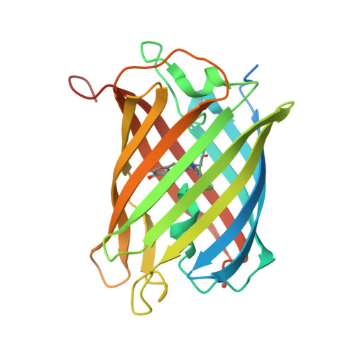
Electrostatic control of photoisomerization pathways in proteins.

Romei, M.G.Lin, C.Y.Mathews, I.I.Boxer, S.G.

(2020) Science 367: 76-79

  • DOI: https://doi.org/10.1126/science.aax1898
  • Primary Citation of Related Structures:  
    6NQJ, 6NQK, 6NQL, 6NQN, 6NQO, 6NQP, 6NQQ, 6NQR, 6NQS, 6NQV

  • PubMed Abstract: 

    Rotation around a specific bond after photoexcitation is central to vision and emerging opportunities in optogenetics, super-resolution microscopy, and photoactive molecular devices. Competing roles for steric and electrostatic effects that govern bond-specific photoisomerization have been widely discussed, the latter originating from chromophore charge transfer upon excitation. We systematically altered the electrostatic properties of the green fluorescent protein chromophore in a photoswitchable variant, Dronpa2, using amber suppression to introduce electron-donating and electron-withdrawing groups to the phenolate ring. Through analysis of the absorption (color), fluorescence quantum yield, and energy barriers to ground- and excited-state isomerization, we evaluate the contributions of sterics and electrostatics quantitatively and demonstrate how electrostatic effects bias the pathway of chromophore photoisomerization, leading to a generalized framework to guide protein design.


  • Organizational Affiliation

    Department of Chemistry, Stanford University, Stanford, CA 94305, USA. sboxer@stanford.edu.


Macromolecules
Find similar proteins by:  (by identity cutoff)  |  3D Structure
Entity ID: 1
MoleculeChains Sequence LengthOrganismDetailsImage
Fluorescent protein Dronpa
A, B, C, D, E
A, B, C, D, E, F, G, H
255Echinophyllia sp. SC22Mutation(s): 3 
Gene Names: Dronpa
UniProt
Find proteins for Q5TLG6 (Echinophyllia sp. SC22)
Explore Q5TLG6 
Go to UniProtKB:  Q5TLG6
Entity Groups  
Sequence Clusters30% Identity50% Identity70% Identity90% Identity95% Identity100% Identity
UniProt GroupQ5TLG6
Sequence Annotations
Expand
  • Reference Sequence
Small Molecules
Modified Residues  1 Unique
IDChains TypeFormula2D DiagramParent
KZV
Query on KZV
A, B, C, D, E
A, B, C, D, E, F, G, H
L-PEPTIDE LINKINGC14 H14 F N3 O4 SCYS, TYR, GLY
Experimental Data & Validation

Experimental Data

  • Method: X-RAY DIFFRACTION
  • Resolution: 2.00 Å
  • R-Value Free: 0.222 
  • R-Value Work: 0.195 
  • R-Value Observed: 0.196 
  • Space Group: P 1 21 1
Unit Cell:
Length ( Å )Angle ( ˚ )
a = 81.75α = 90
b = 86.48β = 94.89
c = 143.81γ = 90
Software Package:
Software NamePurpose
PHENIXrefinement
XDSdata reduction
XDSdata scaling
PHENIXphasing

Structure Validation

View Full Validation Report


Entry History & Funding Information

Deposition Data


Funding OrganizationLocationGrant Number
National Institutes of Health/National Institute of General Medical Sciences (NIH/NIGMS)United States27738
National Institutes of Health/National Institute of General Medical Sciences (NIH/NIGMS)United States118044

Revision History  (Full details and data files)

  • Version 1.0: 2019-06-12
    Type: Initial release
  • Version 1.1: 2019-12-25
    Changes: Database references
  • Version 1.2: 2020-01-01
    Changes: Author supporting evidence
  • Version 1.3: 2020-01-15
    Changes: Database references
  • Version 1.4: 2020-02-05
    Changes: Database references
  • Version 1.5: 2023-10-11
    Changes: Data collection, Database references, Refinement description, Structure summary
  • Version 2.0: 2023-11-15
    Changes: Atomic model, Data collection, Derived calculations