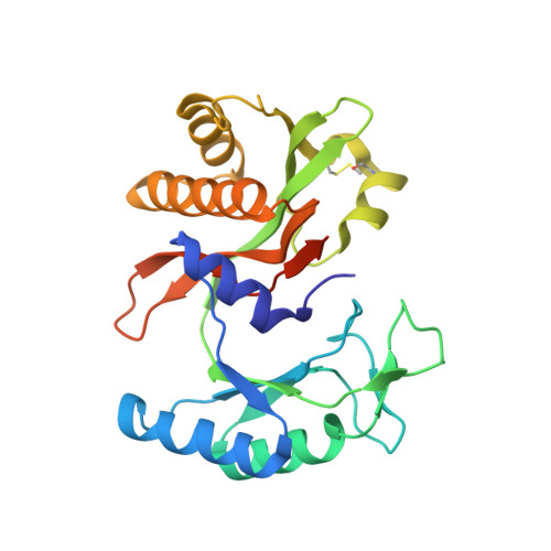
ATP-specificity of succinyl-CoA synthetase from Blastocystis hominis.
Huang, J., Nguyen, V.H., Hamblin, K.A., Maytum, R., van der Giezen, M., Fraser, M.E.(2019) Acta Crystallogr D Struct Biol 75: 647-659
- PubMed: 31282474
- DOI: https://doi.org/10.1107/S2059798319007976
- Primary Citation of Related Structures:
6NO0, 6NO1, 6NO2, 6NO3, 6NO4, 6NO5, 6NO6 - PubMed Abstract:
Succinyl-CoA synthetase (SCS) catalyzes the only step of the tricarboxylic acid cycle that leads to substrate-level phosphorylation. Some forms of SCS are specific for ADP/ATP or for GDP/GTP, while others can bind all of these nucleotides, generally with different affinities. The theory of `gatekeeper' residues has been proposed to explain the nucleotide-specificity. Gatekeeper residues lie outside the binding site and create specific electrostatic interactions with incoming nucleotides to determine whether the nucleotides can enter the binding site. To test this theory, the crystal structure of the nucleotide-binding domain in complex with Mg 2+ -ADP was determined, as well as the structures of four proteins with single mutations, K46βE, K114βD, V113βL and L227βF, and one with two mutations, K46βE/K114βD. The crystal structures show that the enzyme is specific for ADP/ATP because of interactions between the nucleotide and the binding site. Nucleotide-specificity is provided by hydrogen-bonding interactions between the adenine base and Gln20β, Gly111β and Val113β. The O atom of the side chain of Gln20β interacts with N6 of ADP, while the side-chain N atom interacts with the carbonyl O atom of Gly111β. It is the different conformations of the backbone at Gln20β, of the side chain of Gln20β and of the linker that make the enzyme ATP-specific. This linker connects the two subdomains of the ATP-grasp fold and interacts differently with adenine and guanine bases. The mutant proteins have similar conformations, although the L227βF mutant shows structural changes that disrupt the binding site for the magnesium ion. Although the K46βE/K114βD double mutant of Blastocystis hominis SCS binds GTP better than ATP according to kinetic assays, only the complex with Mg 2+ -ADP was obtained.
Organizational Affiliation:
Department of Biological Sciences, University of Calgary, 2500 University Drive NW, Calgary, Alberta T2N 1N4, Canada.